Search Journal-type in search term and press enter
Southwest Pulmonary and Critical Care Fellowships
In Memoriam
Social Media

Critical Care

Last 50 Critical Care Postings

(Most recent listed first. Click on title to be directed to the manuscript.)

May 2025 Critical Care Case of the Month: Where’s the Rub?
April 2025 Critical Care Case of the Month: Being Decisive During a
   Difficult Treatment Dilemma 
January 2025 Critical Care Case of the Month: A 35-Year-Old Admitted After
   a Fall
October 2024 Critical Care Case of the Month: Respiratory Failure in a
   Patient with Ulcerative Colitis
July 2024 Critical Care Case of the Month: Community-Acquired
   Meningitis
April 2024 Critical Care Case of the Month: A 53-year-old Man Presenting
   with Fatal Acute Intracranial Hemorrhage and Cryptogenic Disseminated
   Intravascular Coagulopathy 
Delineating Gastrointestinal Dysfunction Variants in Severe Burn Injury
   Cases: A Retrospective Case Series with Literature Review
Doggonit! A Classic Case of Severe Capnocytophaga canimorsus Sepsis
January 2024 Critical Care Case of the Month: I See Tacoma
October 2023 Critical Care Case of the Month: Multi-Drug Resistant
   K. pneumoniae
May 2023 Critical Care Case of the Month: Not a Humerus Case
Essentials of Airway Management: The Best Tools and Positioning for 
   First-Attempt Intubation Success (Review)
March 2023 Critical Care Case of the Month: A Bad Egg
The Effect of Low Dose Dexamethasone on the Reduction of Hypoxaemia
   and Fat Embolism Syndrome After Long Bone Fractures
Unintended Consequence of Jesse’s Law in Arizona Critical Care Medicine
Impact of Cytomegalovirus DNAemia Below the Lower Limit of
   Quantification: Impact of Multistate Model in Lung Transplant Recipients
October 2022 Critical Care Case of the Month: A Middle-Aged Couple “Not
   Acting Right”
Point-of-Care Ultrasound and Right Ventricular Strain: Utility in the
   Diagnosis of Pulmonary Embolism
Point of Care Ultrasound Utility in the Setting of Chest Pain: A Case of
   Takotsubo Cardiomyopathy
A Case of Brugada Phenocopy in Adrenal Insufficiency-Related Pericarditis
Effect Of Exogenous Melatonin on the Incidence of Delirium and Its 
   Association with Severity of Illness in Postoperative Surgical ICU Patients
Pediculosis As a Possible Contributor to Community-Acquired MRSA
   Bacteremia and Native Mitral Valve Endocarditis
April 2022 Critical Care Case of the Month: Bullous Skin Lesions in
   the ICU
Leadership in Action: A Student-Run Designated Emphasis in
   Healthcare Leadership
MSSA Pericarditis in a Patient with Systemic Lupus
   Erythematosus Flare
January 2022 Critical Care Case of the Month: Ataque Isquémico
   Transitorio in Spanish 
Rapidly Fatal COVID-19-associated Acute Necrotizing
Encephalopathy in a Previously Healthy 26-year-old Man 
Utility of Endobronchial Valves in a Patient with Bronchopleural Fistula in
   the Setting of COVID-19 Infection: A Case Report and Brief Review
October 2021 Critical Care Case of the Month: Unexpected Post-
   Operative Shock 
Impact of In Situ Education on Management of Cardiac Arrest after
   Cardiac Surgery
A Case and Brief Review of Bilious Ascites and Abdominal Compartment
   Syndrome from Pancreatitis-Induced Post-Roux-En-Y Gastric Remnant
   Leak
Methylene Blue Treatment of Pediatric Patients in the Cardiovascular
   Intensive Care Unit
July 2021 Critical Care Case of the Month: When a Chronic Disease
   Becomes Acute
Arizona Hospitals and Health Systems’ Statewide Collaboration Producing a
   Triage Protocol During the COVID-19 Pandemic
Ultrasound for Critical Care Physicians: Sometimes It’s Better to Be Lucky
   than Smart
High Volume Plasma Exchange in Acute Liver Failure: A Brief Review
April 2021 Critical Care Case of the Month: Abnormal Acid-Base Balance
   in a Post-Partum Woman
First-Attempt Endotracheal Intubation Success Rate Using A Telescoping
   Steel Bougie
January 2021 Critical Care Case of the Month: A 35-Year-Old Man Found
   Down on the Street
A Case of Athabaskan Brainstem Dysgenesis Syndrome and RSV
   Respiratory Failure
October 2020 Critical Care Case of the Month: Unexplained
   Encephalopathy Following Elective Plastic Surgery
Acute Type A Aortic Dissection in a Young Weightlifter: A Case Study with
  an In-Depth Literature Review
July 2020 Critical Care Case of the Month: Not the Pearl You Were
   Looking For...
Choosing Among Unproven Therapies for the Treatment of Life-Threatening
   COVID-19 Infection: A Clinician’s Opinion from the Bedside
April 2020 Critical Care Case of the Month: Another Emerging Cause
   for Infiltrative Lung Abnormalities
Further COVID-19 Infection Control and Management Recommendations for
   the ICU
COVID-19 Prevention and Control Recommendations for the ICU
Loperamide Abuse: A Case Report and Brief Review
Single-Use Telescopic Bougie: Case Series


For complete critical care listings click here.

The Southwest Journal of Pulmonary and Critical Care publishes articles directed to those who treat patients in the ICU, CCU and SICU including chest physicians, surgeons, pediatricians, pharmacists/pharmacologists, anesthesiologists, critical care nurses, and other healthcare professionals. Manuscripts may be either basic or clinical original investigations or review articles. Potential authors of review articles are encouraged to contact the editors before submission, however, unsolicited review articles will be considered.

------------------------------------------------------------------------------------

Wednesday
Jan012020

January 2020 Critical Care Case of the Month: A Code Post Lung Needle Biopsy

Sarika Savajiyani MD and Clement U. Singarajah MBBS

 Phoenix VA Medical Center

Phoenix, AZ USA


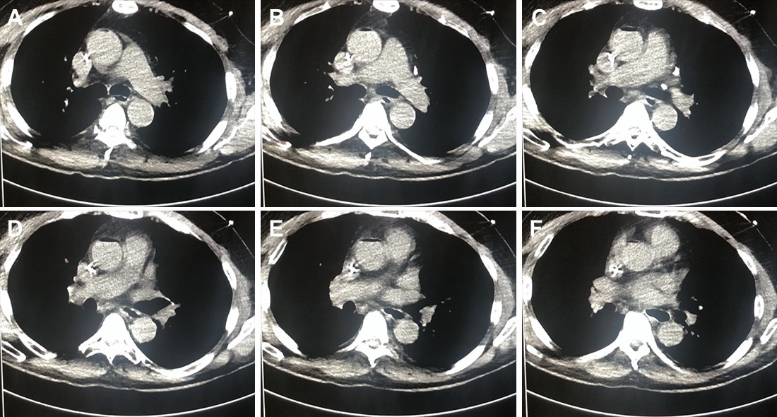
A 67-year-old man with a history of stage IIA rectal adenocarcinoma post neoadjuvant chemoradiation presented with a near code event after elective CT guided biopsy of an enlarging left lower lobe lung nodule. The patient became bradycardic and profoundly hypotensive immediately after the CT guided biopsy with the following vital signs: Systolic BP < 90 mmHg, HR 40/min sinus bradycardia, SpO2 on 100% oxygen non rebreather was 90%. Telemetry and EKG showed ST elevation in the anterior leads. He complained of vague arm and leg weakness and tingling, but did not lose consciousness or suffer a cardiac arrest. 

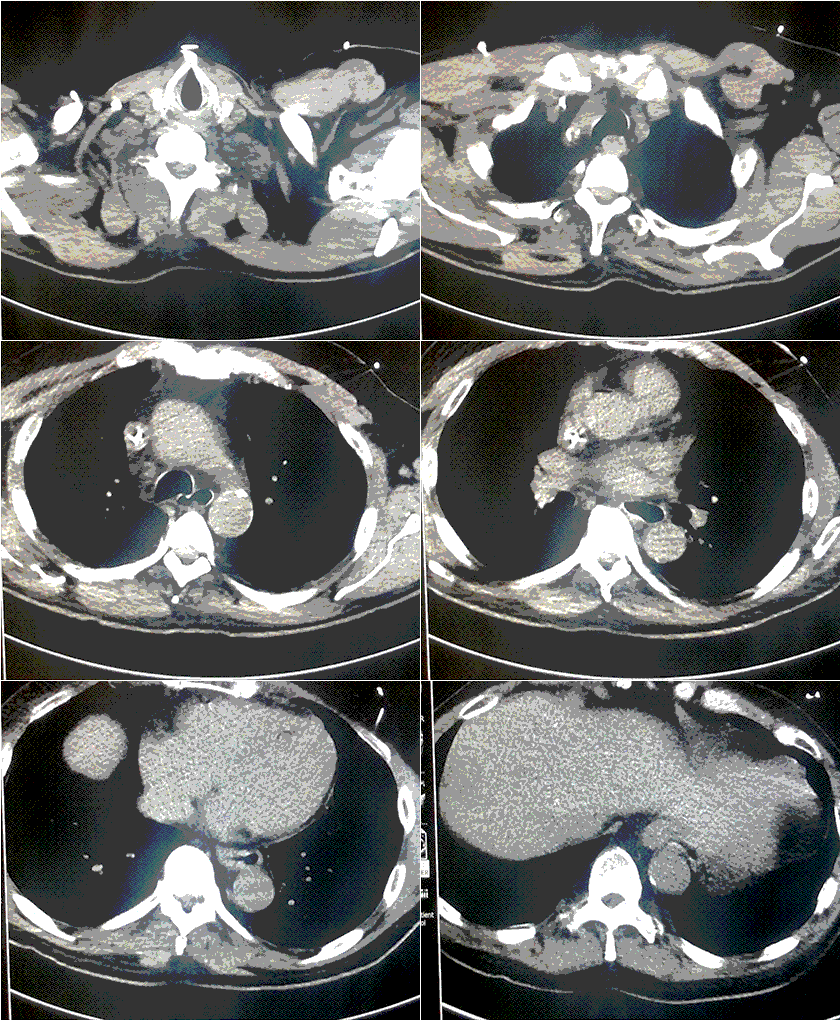
A CT scan was performed about 2-3 minutes after the patient deteriorated (Figure 1).

Figure 1. A-E: Representative images from CT scan in soft tissue windows. Lower: Video of CT scan in soft tissue windows.

What radiographic finding likely explains the patient’s clinical deterioration?  (Click on the correct answer to be directed to the second of six pages)

Cite as: Savajiyani S, Singarajah CU. January 2020 critical care case of the month: a code post lung needle biopsy. Southwest J Pulm Crit Care. 2020;20(1):1-6. doi: https://doi.org/10.13175/swjpcc042-19 PDF

Tuesday
Oct012019

October 2019 Critical Care Case of the Month: Running Naked in the Park

Spencer Jasper MD

Matthew Adams DO

Jonathan Boyd MD

Jeremiah Garrison MD

Janet Campion MD

The University of Arizona College of Medicine

Tucson, AZ USA


History of Present Illness

A 34-year-old man with a history of IV drug abuse was brought into emergency department by EMS and Tucson Police Department after complaints of naked man running and behaving erratically in a park. On arrival to emergency department patient was acting aggressively towards staff, spitting and attempting to bite. The ER staff attempted multiple times to sedate the patient with benzodiazepines, however, due to continued aggressive behavior, ongoing encephalopathy and the need for increased sedation, the patient was intubated for airway protection.

The patient was febrile (40.6° C), tachycardic (122) and hypertensive (143/86). On physical exam patient was not cooperative, was diaphoretic, cachectic, with reactive constrictive pupils, track marks in antecubital fossa bilaterally. No clonus or hypertonicity. During intubation, there was noted to be nuchal rigidity.

He was then admitted to the medical ICU. Drug intoxication from possible methamphetamines was the presumptive diagnosis of encephalopathy but given nuchal rigidity and fevers there was concern for other etiologies.

Physical Exam

  • Vitals: T 40.6 °C, HR: 122, RR: 22, BP: 143/86, SpO2: 97% WT: 55 kg
  • General: Intubated and sedated, cachectic
  • Eye: Pupils constricted but reactive to light
  • HEENT: Normocephalic, atraumatic
  • Neck: Stiff, non-tender, no carotid bruits, no JVD, no lymphadenopathy
  • Lungs: Clear to auscultation, non-labored respiration
  • Heart: Normal rate, regular rhythm, no murmur, gallop or peripheral edema
  • Abdomen: Soft, non-tender, non-distended, normal bowel sounds, no masses
  • Skin: Skin is warm, dry and pink, multiple abrasions on the lower extremities bilaterally, track marks noted in the antecubital fossa bilaterally. Large abrasion with bruising around the right knee and erythema and welts on the right shin. Erythematous area on the dorsal surface of the right hand
  • Neurologic: Nonfocal prior to intubation, no clonus or hypertonicity noted

Drug overdose/intoxication was presumptive diagnosis for his acute encephalopathy. Based on physical exam and vitals, what other etiologies should be considered? (click on the correct answer to be directed to the second of seven pages)

  1. Embolic stroke
  2. Heat stroke
  3. Hyperthyroidism
  4. Meningitis
  5. All of the above

Cite as: Jasper S, Adams M, Boyd J, Garrison J, Campion J. October 2019 critical care case of the month: running naked in the park. Southwest J Pulm Crit Care. 2019;19(4):110-8. doi: https://doi.org/10.13175/swjpcc054-19 PDF

Wednesday
Aug212019

Severe Accidental Hypothermia in Phoenix? Active Rewarming Using Thoracic Lavage

Michael Mozer BS1

Guy Raz, MD2

Ryan Wyatt, MD2

Alexander Toledo, DO, PharmD2

1University of New England College of Osteopathic Medicine

Biddeford, ME USA

2Department of Emergency Medicine

Maricopa Medical Center, Phoenix, AZ USA


Abstract

Hypothermia can progress quickly and become life threatening if left untreated. Rewarming in the severely hypothermic patient is of critical importance and is achieved with active and passive techniques. Here we present a case of a hypothermic patient with cardiac instability for whom thoracic lavage was ultimately used. We will review the treatment of hypothermia and discuss the technical aspects our approach.

Case Presentation

A 53 year-old male with a past medical history of substance abuse, chronic hepatitis C, and poorly controlled type 2 diabetes mellitus complicated by a recent hospitalization for osteomyelitis was brought to the emergency department after being found lying on a road in a shallow pool of water in the early morning hours of a rainy day in Phoenix, Arizona. The ambient temperature that night was 39 °F (3.9 °C). Emergency Medical Services (EMS) noted a decreased level of consciousness and obtained a finger stick glucose of 15 mg/dl. EMS reported a tympanic membrane temperature of 23.9 °C. En route, the patient was administered 2mg naloxone and 25g dextrose intravenously with no improvement in his mental status. On Emergency Department (ED) arrival, the patient had a GCS of 8 (Eyes 4, Verbal 1, Motor 3) and exhibited intermittent posturing. His foot wound appeared clean and without signs of infection. The initial core temperature recorded was 25.9°C via bladder thermometer, systolic blood pressure was 92/50, and heart rate fluctuated between 50 and 90 beats per minute.

After removing wet clothing, initiation of warmed saline, and placing a forced warm air blanket on the patient, he was intubated for airway protection and vasopressors were initiated. Osborn waves were evident on the initial EKG (Figure 1).

Figure 1. Initial EKG with Osborn Waves (arrows).

A warmed ventilator circuit was initiated with only 0.5 °C increase in temperature in first 30 minutes. Despite these measures, he remained hypotensive and unstable. Significant laboratory findings were a white blood cell count of 25.5 thousand (92% neutrophils), lactic acid of 7.6, potassium of 5.8, serum creatinine of 1.05, glucose of 283, INR of 1.1, and urine drug screen positive for cocaine. Given his recalcitrance to norepinephrine and risk of death secondary to fatal dysrhythmia with temperatures below 28 °C intrathoracic lavage initiated.

The right hemithorax was selected for irrigation because left-sided tube placement can induce ventricular fibrillation in a perfusing patient (1). Using standard sterile technique, two 36 French thoracostomy tubes were placed; the first in the second intercostal space along the mid-clavicular line, and the second in the 5th intercostal space in the posterior axillary line (1-3). The tips of the thoracostomy tubes were oriented such that the anterior-superior tube was positioned near the right apex and the lateral-inferior tip was positioned low in the thoracic cavity (1,3). To maintain the temperature of the instilled fluid, a fluid warmer system (Level 1; Smiths Medical; Minneapolis, MN) was used and set to 41 °C. A Christmas tree adapter was used to connect the IV tubing to the superior thoracostomy tube, and a water seal chamber was attached to the inferior tube for passive drainage (3). Flow through the system was targeted to maintain steady passive drainage as described in the literature (1-6).

Thoracic cavity lavage with 41 °C saline was performed and the patient was transferred to the medical ICU after 3 hours in the ED. When he was transferred his core temperature was 29 °C and he remained on norepinephrine for hemodynamic instability. After 2 hours of continued rewarming in the MICU, his core temperature was 32 °C. Osborn waves evident on initial EKG were resolved (Figure 2).

Figure 2. Repeat EKG showing resolution of Osborn waves.

The patient left against medical advice from the hospital 4 days later neurologically intact and without sequela.

Discussion

Hypothermia can be clinically classified as mild, moderate or severe (7). Mild hypothermia, defined as core temperatures of 32-35 °C, presents with shivering. Amnesia, dysarthria, ataxia, tachycardia, and tachypnea can also be seen (1). Moderate hypothermia, defined as core temperatures of 28-32 °C, usually can present with or without shivering. Stupor, hypoventilation, paradoxical undressing and non-fatal arrhythmias such as atrial fibrillation and junctional bradycardia may also be seen (1). Patients with severe hypothermia, generally defined as temperatures below 28 °C, can present with coma, areflexia, pulmonary edema, bradycardia, and hypotension (1). There is a significant risk of fatal cardiac dysrhythmias without rapid therapeutic rewarming (1,7,8).

Rewarming in the hypothermic patient is of critical importance and is achieved with passive and/or active techniques. Attempts to limit heat loss are often unsuccessful, especially in the absence of a normal shiver response. It however remains as the first line treatment for hypothermia (8-10). Passive rewarming is attempted by the removal of cold/wet clothing and keeping the patient covered (8-10). Active external rewarming (AER) is the next line of treatment and consist of the use of externally rewarming devices such as warmed blankets, warm environment, forced air warming (Bair Hugger; 3M; Maplewood, MN) or warm hot water bladders placed in the groin and axilla (1,7-10). Active Internal Rewarming (AIR) techniques can be used to achieve more rapid increases in core temperature and are primarily utilized in cases of cardiac instability or if AER is unsuccessful (8). When available, the method of choice for active internal rewarming (AIR) is cardiopulmonary bypass (CPB) or extracorporeal membrane oxygenation (ECMO) as they can achieve the fastest increase in core temperature (9 °C/hr and 6 °C/hr respectively) and provide cardiovascular support (1,8,11,12). Several techniques are described in the literature that can be considered if CPB or ECMO are unavailable. These include esophageal warming devices, endovascular catheters, hemodialysis, and endocavitary lavage (1,2,4-6,13-15). While no randomized controlled trials exist, several case reports and reviews have tried to compare various techniques. These sources to do not seem to favor any particular technique over another but rather reports the rates of temperature rise (1-3,5-7,13-15). Classically, lavage techniques are attempted in the thoracic cavity, the peritoneum, the bladder, the stomach, the esophagus, or the colon. These techniques are generally coupled with warm IV fluids and warming air through the ventilator to limit loss of body heat to iatrogenic procedures during the rewarming attempt (1,7). Thoracic lavage is effective with a reported rewarming rates of 3-6 °C/hr and with excellent outcomes in case reports (1,2,4-6). Here we present a case of a hypothermic patient with cardiac instability where thoracic lavage is used and discuss the technical aspects of this approach.

Our case demonstrates the efficacy of utilizing thoracic cavity lavage for rapid rewarming in patients with severe hypothermia with a pulse and at high risk of fatal cardiac arrhythmia. In multiple case reports, thoracic lavage has been used successfully in hypothermic patients who suffered complete cardiopulmonary collapse requiring CPR (2,4,5). Although warm thoracic lavage is not the treatment of choice in these circumstances, in a facility not equipped with ECMO or CPB and a patient too unstable to transfer, it seemed to us to be the best technique. Gastric, colonic, and bladder lavage offer very minimal heat transfer due to limitations in surface area (2).

Hemodialysis would have required for us to call in a technician and would have required approval by a nephrologist at our institution. Available central venous rewarming catheters require bypass of a failsafe mechanism that does not allow rewarming to be initiated below 30 °C (1). Peritoneal lavage was a plausible choice but does not directly warm the mediastinum (2). While an open mediastinal technique has been used, we did not feel it was appropriate in a patient with a concurrent pulse (1,3). Thoracic lavage is therefore an effective alternative that should be used in cases where CPB and ECMO are unavailable especially in a patient that is hemodynamically unstable and may not survive transfer. The equipment is readily available to any Emergency Medicine or Critical Care physician. In addition, this case exemplifies the positive outcomes that are associated with rapid rewarming in the hypothermic patient with a pulse. We believe our case demonstrates the efficacy of this technique for myocardial protection from hemodynamic collapse, a topic which has not been adequately studied in the literature.

References

  1. Brown DJ, Danzl DF. Accidental hypothermia. In: Auerbach PS, ed. Wilderness Medicine. 7th ed. St. Louis: Mosby Inc.; 2017:135-62.
  2. Plaisier BR. Thoracic lavage in accidental hypothermia with cardiac arrest--report of a case and review of the literature. Resuscitation. 2005 ;66(1):99-104. [CrossRef] [PubMed]
  3. Schiebout JD. Hypothermic Patient Management. In: Reichman EF. eds. Reichman's Emergency Medicine Procedures, 3e New York, NY: McGraw-Hill. Available at: http://accessemergencymedicine.mhmedical.com/content.aspx?bookid=2498&sectionid=201303754 (accessed August 02, 2019).
  4. Little G. Accidental hypothermic cardiac arrest and rapid mediastinal warming with pleural lavage: A survivor after 3.5 hours of manual CPR. BMJ Case Reports. July 2017:bcr-2017-220900. [CrossRef] [PubMed]
  5. Turtiainen J, Halonen J, Syväoja S, Hakala T. Rewarming a patient with accidental hypothermia and cardiac arrest using thoracic lavage. Ann Thorac Surg. 2014 Jun;97(6):2165-6. [CrossRef] [PubMed]
  6. Ellis MM, Welch RD. Severe hypothermia and cardiac arrest successfully treated without external mechanical circulatory support. Am J Emerg Med. 2016;34(9):1913.e5-6. [CrossRef] [PubMed]
  7. Tintinalli J, Stapczynski J, Ma O, Yealy D, Meckler G, Cline D. Tintinalli's Emergency Medicine. 8th ed. New York, NY: McGraw-Hill Education; 2016:1743-4.
  8. Brugger H, Boyd J, Paal P. Accidental Hypothermia. N Engl J Med. 2012;367(20):1930-8. [CrossRef] [PubMed]
  9. Paal P, Gordon L, Strapazzon G, et al. Accidental hypothermia-an update: The content of this review is endorsed by the International Commission for Mountain Emergency Medicine (ICAR MEDCOM). Scand J Trauma Resusc Emerg Med. 2016;24(1):111. [CrossRef] [PubMed]
  10. Zafren K, Giesbrecht GG, Danzl DF, et al. Wilderness Medical Society practice guidelines for the out-of-hospital evaluation and treatment of accidental hypothermia: 2014 update. Wilderness Environ Med. 2014 Dec;25(4 Suppl):S66-85. [CrossRef] [PubMed]
  11. Schober A, Sterz F, Handler C, et al. Cardiac arrest due to accidental hypothermia-A 20 year review of a rare condition in an urban area. Resuscitation. 2014;85(6):749-56. [CrossRef] [PubMed]
  12. Saczkowski RS, Brown DJA, Abu-Laban RB, Fradet G, Schulze CJ, Kuzak ND. Prediction and risk stratification of survival in accidental hypothermia requiring extracorporeal life support: An individual patient data meta-analysis. Resuscitation. 2018;127:51-7.[CrossRef] [PubMed]
  13. Primozic KK, Svensek F, Markota A, Sinkovic A. Rewarming after severe accidental hypothermia using the esophageal heat transfer device: a case report. Ther Hypothermia Temp Manag. 2018 Mar;8(1):62-4. [CrossRef] [PubMed]
  14. Murakami T, Yoshida T, Kurokochi A, et al. Accidental hypothermia treated by hemodialysis in the acute phase: three case reports and a review of the literature. Intern Med. 2019 Jun 7. [CrossRef]
  15. Klein LR, Huelster J, Adil U, et al. Endovascular rewarming in the emergency department for moderate to severe accidental hypothermia. Am J Emerg Med. 2017 Nov;35(11):1624-9. [CrossRef] [PubMed]

Cite as: Mozer M, Raz G, Wyatt R, Toledo A. Severe accidental hypothermia in Phoenix? Active rewarming using thoracic lavage. Southwest J Pulm Crit Care. 2019;19(2):79-83. doi: https://doi.org/10.13175/swjpcc038-19 PDF 

Wednesday
Aug142019

Left Ventricular Assist Devices: A Brief Overview

Bhargavi Gali MD

Department of Anesthesiology and Perioperative Medicine

Division of Critical Care Medicine

Mayo Clinic Minnesota

Rochester, MN, USA


Introduction

Second and third generation left ventricular assist devices (LVAD) have been increasingly utilized as both a bridge to transplantation and as destination therapy (in patients who are not considered transplant candidates) for advanced heart failure. Currently approximately 2500 LVADs are implanted yearly, with an estimated one year survival of >80% (1). Almost half of these patients undergo implantation as destination therapy. A recent systematic review and meta-analysis found no difference in one-year mortality between patients undergoing heart transplantation in comparison with patients undergoing LVAD placement (2).

Early LVADs were pulsatile pumps, but had multiple limitations including duration of device function, and requirement for a large external lead that increased risk of infection. Currently utilized second and third generation devices are continuous flow (first generation were pulsatile flow). Second generation devices have axial pumps (HeartMate II®). The third generation LVADs ((HeartMate III®), HVAD®) are also continuous flow, with centrifugal pumps, which are thought to decrease possibility of thrombus formation and increase pump duration in comparison to the second generation axial pumps. It is also felt that a lack of mechanical bearings contributes to this effect.

LVADs support circulation by either replacing or supplementing cardiac output. Blood is drained from the left ventricle with inflow cannula in the left ventricular apex to the pump, and blood is returned to the ascending aorta via the outflow cannula (3) (Figure 1).

Figure 1. Third generation Left Ventricular Assist Device. Heartware System ™. Continuous flow left ventricular assist device (LVAD) configuration. One of the third generation LVADs is the HeartWare System. With this device the inflow cannula is integrated into the pump. The pump is attached to the heart in the pericardial space, with the outflow cannula in the aorta. A driveline connects the device to the control unit. This control unit is attached to the two batteries. (Figure used with permission from Medtronic).

The device assists the left ventricle by the action of the axial (second generation) or centrifugal (third generation) pump that rotates at a very high speed and ejects the blood into the aorta via the outflow cannula. A tunneled driveline connects the pump to the external controller that operates the pump function. The controller connects to the power source via two cables, which can be battery or module-powered.

LVADs offload volume from the left ventricle, and decrease left ventricular work. Pulmonary pressures and the trans pulmonary gradients are also decreased by the reduced volume in the left ventricle (4). End organ perfusion is improved secondary to enhanced arterial blood pressure and micro perfusion.

There are four main parameters of pump function:

  • Pump speed: the speed at which the LVAD rotors spin, and is programmed. Measured in RPM.
  • Pump power: the wattage needed to maintain speed and flow, which is the energy needed to run the pump. Measured in Watts.
  • Pump flow: estimate of the cardiac output, which is the blood returned to the ascending aorta, and is based on pump speed and power. Measure in L/min
  • Pulsatility index (PI): a calculated value that indicates assistance the pump provides, in relation to intrinsic left ventricular A higher number indicates higher left ventricular contribution to pulsatile flow.

The cardiac output of currently utilized LVADs is directly related to pump speed and inversely related to the pressure gradient across the pump. As the pump speed is fixed, right ventricular failure can decrease the volume of blood transmitted to the pump and decrease LVAD flow (3, 4). With right ventricular failure, inotropic support may be needed to improve the LVAD pump flow. High afterload, such as may be seen with an increase in systemic vascular resistance can decrease pump flow.

Complications

Adverse events occur in more than 70% of LVAD patients in the first year (5). These complications include infections, bleeding, stroke, and LVAD thrombosis. More than 50% of patients are readmitted within the first 6 months after LVAD implantation (6).

Driveline infections are the most commonly reported LVAD infection, and are the most likely to respond to treatment (7). Pump pocket infections may require debridement plus/minus antibiotic bead placement. Bloodstream infections are less commonly reported, and more difficult to treat, and many patients are placed on chronic suppressive antibiotic therapy (7). There is a possible association between stroke and bloodstream infection, reported in some studies. Patients who were younger and had a higher body mass index were noted to have a higher incidence of LVAD infections.

Gastrointestinal bleeding is a major cause of nonsurgical bleeding, reported in almost 30% of patients after LVAD placement (1). Patients may develop acquired von Willebrand factor deficiency secondary to high shear forces in the LVAD that lead to breakdown of von Willebrand protein (6). Antithrombotic therapy is commonly instituted after LVAD implantation which also increases risk of bleeding. A high incidence of arteriovenous malformations is reported in these patients, although the etiology is not clear. Transfusion, holding antithrombotic therapy, and identifying possible sources are included in the standard approach to management.

There is a high risk of both ischemic and hemorrhagic strokes in the first year after LVAD placement (8). Surgical closure of the aortic valve and off-axis positioning of the cannulas have been suggested as altering shear forces, increasing thrombotic risk, and thus risk of stroke.  Post-surgical risks may include pump thrombosis, infections, supratherapeutic INR, and poorly controlled hypertension. Early diagnosis has led to consideration of interventions such as thrombectomy (8).

LVAD thrombosis can occur on either cannula (inflow or outflow) or the pump. Typically patients receive ongoing anticoagulation, commonly with warfarin, and antiplatelet agents, and often aspirin. Heartmate II® may have higher rate of thrombosis than HVAD or Heart Mate 3, although this is under debate (6). Thrombotic complications range in severity from asymptomatic increase in lactate dehydrogenase or plasma-free hemoglobin, to triggering of LVAD alarms, up to development of heart failure. The inflow and outflow cannulas and pump can be the site of thrombosis. Management typically involves revising the antithrombotic management. If there is no improvement or worsening, replacement of LVAD may be indicated. There is limited evidence to suggest that systemic thrombolysis may be of benefit in treating pump thrombosis, particularly in regards to the HVAD, though better data would be useful

Procedural Management

When a patient with an LVAD requires non cardiac surgery, optimal management includes having an on-site VAD technician, and close involvement of VAD cardiology and cardiac surgery in consultation. Anticoagulation will often be transitioned to heparin infusion prior to elective procedures (9). Suction events (LV wall is sucked into the inflow cannula) can occur secondary to under filled left heart, and this can become more apparent perioperatively. This can also decrease right heart contractility by moving the interventricular septum to the left, and thus decrease cardiac output. Management often involves fluid bolus. Suction events can lead to decreased flow, left ventricular damage, and ventricular arrhythmias. Hemodynamic management can be challenging with non-pulsatile flow, and placement of an arterial line can facilitate optimal management. Postoperative care in a monitored setting is beneficial in case of further volume related events and to watch for bleeding.

Emergent Complications

Arrhythmias occur in many patients after LVAD implantation. Atrial arrhythmias are reported in up to half of LVAD patients, and ventricular arrhythmias in 22-59% (10, 11).  Loss of AV synchrony can lead to decreased LV filling and subsequent RV failure. Rhythm or rate control with rapid atrial arrhythmias is necessary to decrease development of heart failure. Ventricular arrhythmias may be hemodynamically tolerated for some time secondary to the LVAD support (6).  If there is concern that the inflow cannula is touching the LV septum, as may occur with severe hypovolemia, echocardiography can help determine if volume resuscitation should be the initial step in treating ventricular arrhythmia.

If cardiac arrest occurs, the first step of cardiopulmonary resuscitation in patients with LVAD is assessment of appropriate perfusion via physical examination (12). If perfusion is poor or absent, assessment of LVAD function should take place. If the LVAD is not functioning appropriately, checking for connections and power is the next step. If unable to confirm function or restart LVAD, chest compressions are indicated by most recent guidelines from the American Heart Association. There is always concern of dislodgement of LVAD cannula or bleeding during these situations.

Conclusion

Currently implanted LVADS are continuous flow, and provide support via a parallel path from the left ventricle to the aorta. As the number of patients with LVADs increase all medical providers should have a basic understanding of the function and common complications associated with these devices. This will enhance the ability to initiate appropriate care.

References

  1. Kirklin JK, Pagani FD, Kormos RL, et al. Eighth annual INTERMACS report: Special focus on framing the impact of adverse events. J Heart Lung Transplant. 2017 Oct;36(10):1080-6. [CrossRef] [PubMed]
  2. Theochari CA, Michalopoulos G, Oikonomou EK, et al. Heart transplantation versus left ventricular assist devices as destination therapy or bridge to transplantation for 1-year mortality: a systematic review and meta-analysis. Annals of Cardiothoracic Surgery. 2017;7(1):3-11. [CrossRef] [PubMed]
  3. Lim HS, Howell N, Ranasinghe A. The physiology of continuous-flow left ventricular assist devices. J Card Fail. 2017;23(2):169-80. [CrossRef] [PubMed]
  4. Roberts SM, Hovord DG, Kodavatiganti R, Sathishkumar S. Ventricular assist devices and non-cardiac surgery. BMC Anesthesiology. 2015;15(1):185. [CrossRef] [PubMed]
  5. Miller LW, Rogers JG. Evolution of left ventricular assist device therapy for advanced heart failure: a review. JAMA Cardiol. 2018 Jul 1;3(7):650-8. [CrossRef] [PubMed]
  6. DeVore AD, Patel PA, Patel CB. Medical management of patients with a left ventricular assist device for the non-left ventricular assist device specialist. JACC Heart Fail. 2017 Sep;5(9):621-31. [CrossRef] [PubMed]
  7. O'Horo JC, Abu Saleh OM, Stulak JM, Wilhelm MP, Baddour LM, Rizwan Sohail M. Left ventricular assist device infections: a systematic review. ASAIO J. 2018 May/Jun;64(3):287-294. [CrossRef] [PubMed]
  8. Goodwin K, Kluis A, Alexy T, John R, Voeller R. Neurological complications associated with left ventricular assist device therapy. pert Rev Cardiovasc Ther. 2018 Dec;16(12):909-17. [CrossRef] [PubMed]
  9. Barbara DW, Wetzel DR, Pulido JN, et al. The perioperative management of patients with left ventricular assist devices undergoing noncardiac surgery. Mayo Clinic Proceedings. 2013;88(7):674-82. [CrossRef] [PubMed]
  10. Enriquez AD, Calenda B, Gandhi PU, Nair AP, Anyanwu AC, Pinney SP. Clinical impact of atrial fibrillation in patients with the heartmate ii left ventricular assist device. J Am Coll Cardiol. 2014 Nov 4;64(18):1883-90. [CrossRef] [PubMed]
  11. Nakahara S, Chien C, Gelow J, et al. Ventricular arrhythmias after left ventricular assist device. Circ Arrhythm Electrophysiol. 2013 Jun;6(3):648-54. [CrossRef] [PubMed]
  12. Peberdy MA, Gluck JA, Ornato JP, et al. Cardiopulmonary resuscitation in adults and children with mechanical circulatory support: a scientific statement from the American Heart Association. Circulation. 2017;135(24):e1115-e34.`[CrossRef] [PubMed]

Cite as: Gali B. Left ventricular assist devices: a brief overview. Southwest J Pulm Crit Care. 2019;19(2):68-72. doi: https://doi.org/10.13175/swjpcc039-19 PDF 

Monday
Jul012019

July 2019 Critical Care Case of The Month: An 18-Year-Old with Presumed Sepsis and Progressive Multisystem Organ Failure 

Robert A. Raschke, MD

The University of Arizona College of Medicine – Phoenix

Phoenix, AZ USA


History of Present Illness

An 18-year-old female student from Flagstaff was transferred to our hospital for refractory sepsis. She had presented with a 2 week history of fever, malaise, sore throat, myalgias, arthralgias and a rash.

PMH, SH and FH

She reported no significant past medical history or family history. She attended cosmetology school, denied smoking or drug abuse and was sexually monogamous. She had only traveled in-state, did not hike or camp and her only animal exposure was playing with her two pet Great Danes.

Physical Examination

The patient had a fever of 38.5°C. on original presentation. HEENT exam was reported as unrevealing. Lungs were clear. There were no heart murmurs and the abdominal exam was unremarkable. No joint effusions were apparent. A rash was mentioned, but not described and it apparently disappeared shortly after admission.

Initial laboratory testing was significant for WBCC of 12.1 K/mm3, creatinine of 1.5 mg/dL and AST of 45 IU/L. A rapid influenza screen, urinalysis and chest radiography were unrevealing. Blood cultures were drawn and intravenous fluids, piperacillin/tazobactam and azithromycin were administered. Over the next four days, the fever persisted and the blood cultures resulted in no growth. Serial laboratory values demonstrated progressive worsening in renal function and increasing hepatic enzymes. The patient became dyspneic and developed rales and progressive hypoxia prompting transfer.

On arrival in our ICU, the patient was alert, in mild respiratory distress and hypotensive to 78/43 mmHg, requiring immediate initiation of intravenous norepinephrine. She reported nausea and severe diffuse myalgia and arthralgia. On examination, she was ill-appearing with blood pressure 101/58 (on norepinephrine at 25 mcg/min), heart rate 104 beats/min, respiratory rate 33 breaths/min, temperature 38.8°C. She had mild oropharyngeal erythema, some shotty cervical lymph nodes, bilateral rales, mild epigastric and right upper quadrant tenderness, and a macular erythematous rash approximately 14 x 29 cm on her left forearm that disappeared within several hours.

Her ICU admission chest x-ray is shown in Figure 1.


Figure 1. Admission ICU portable chest X-ray showing bilateral areas of consolidation.

Her laboratory evaluation showed the following:

  • WBCC: 2,500/mm3 63% segs with toxic granulation/vacuolated segs
  • Hemoglobin/Hematocrit: 7.9 g/dL/26.7%
  • Platelets: 50,000/mm3
  • BUN/creatinine: 23/1.25 mg/dL
  • AST/ALT: 246/189 IU/L (normal 10-40 and 7-56)
  • PT: 20.9 sec
  • Lactate: 4.5 mmol/L
  • Urinalysis: bland sediment, without bacteria or leukocytes
  • ABG: 7.33, pCO2 34, pO2 78 (on 45% FiO2 by ventimask)
  • Transthoracic echocardiogram showed normal LV and RV size and systolic function with no vegetations
  • US abdomen showed hepatosplenomegaly, retroperitoneal lymphadenopathy, and normal kidneys and ureters.

What are diagnostic considerations at this time?  (Click on the correct answer to be directed to the second of six pages)

  1. Rocky mountain spotted fever (RMSF)
  2. Acute retroviral syndrome
  3. Still’s disease
  4. Systemic lupus erythematosus (SLE)
  5. All of the above

Cite as: Raschke RA. July 2019 critical care case of the month: an 18-year-old with presumed sepsis and progressive multisystem organ failure. Southwest J Pulm Crit Care. 2019;19(1):1-9. doi: https://doi.org/10.13175/swjpcc043-19 PDF