Search Journal-type in search term and press enter
Southwest Pulmonary and Critical Care Fellowships
In Memoriam
Social Media

Pulmonary

Last 50 Pulmonary Postings

(Click on title to be directed to posting, most recent listed first)

March 2025 Pulmonary Case of the Month: Interstitial Lung Disease of
   Uncertain Cause
December 2024 Pulmonary Case of the Month: Two Birds in the Bush Is
   Better than One in the Hand
Glucagon‐like Peptide-1 Agonists and Smoking Cessation: A Brief Review
September 2024 Pulmonary Case of the Month: An Ounce of Prevention
   Caused a Pound of Disease
Yield and Complications of Endobronchial Ultrasound Using the Expect
   Endobronchial Ultrasound Needle
June 2024 Pulmonary Case of the Month: A Pneumo-Colic Association
March 2024 Pulmonary Case of the Month: A Nodule of a Different Color
December 2023 Pulmonary Case of the Month: A Budding Pneumonia
September 2023 Pulmonary Case of the Month: A Bone to Pick
A Case of Progressive Bleomycin Lung Toxicity Refractory to Steroid Therapy
June 2023 Pulmonary Case of the Month: An Invisible Disease
February 2023 Pulmonary Case of the Month: SCID-ing to a Diagnosis
December 2022 Pulmonary Case of the Month: New Therapy for Mediastinal
   Disease
Kaposi Sarcoma With Bilateral Chylothorax Responsive to Octreotide
September 2022 Pulmonary Case of the Month: A Sanguinary Case
Electrotonic-Cigarette or Vaping Product Use Associated Lung Injury:
   Diagnosis of Exclusion
June 2022 Pulmonary Case of the Month: A Hard Nut to Crack
March 2022 Pulmonary Case of the Month: A Sore Back Leading to 
   Sore Lungs
Diagnostic Challenges of Acute Eosinophilic Pneumonia Post Naltrexone
Injection Presenting During The COVID-19 Pandemic
Symptomatic Improvement in Cicatricial Pemphigoid of the Trachea
   Achieved with Laser Ablation Bronchoscopy
Payer Coverage of Valley Fever Diagnostic Tests
A Summary of Outpatient Recommendations for COVID-19 Patients
   and Providers December 9, 2021
December 2021 Pulmonary Case of the Month: Interstitial Lung
   Disease with Red Knuckles
Alveolopleural Fistula In COVID-19 Treated with Bronchoscopic 
   Occlusion with a Swan-Ganz Catheter
Repeat Episodes of Massive Hemoptysis Due to an Anomalous Origin 
   of the Right Bronchial Artery in a Patient with a History
   of Coccidioidomycosis
September 2021 Pulmonary Case of the Month: A 45-Year-Old Woman with
   Multiple Lung Cysts
A Case Series of Electronic or Vaping Induced Lung Injury
June 2021 Pulmonary Case of the Month: More Than a Frog in the Throat
March 2021 Pulmonary Case of the Month: Transfer for ECMO Evaluation
Association between Spirometric Parameters and Depressive Symptoms 
   in New Mexico Uranium Workers
A Population-Based Feasibility Study of Occupation and Thoracic
   Malignancies in New Mexico
Adjunctive Effects of Oral Steroids Along with Anti-Tuberculosis Drugs
   in the Management of Cervical Lymph Node Tuberculosis
Respiratory Papillomatosis with Small Cell Carcinoma: Case Report and
   Brief Review
December 2020 Pulmonary Case of the Month: Resurrection or 
   Medical Last Rites?
Results of the SWJPCC Telemedicine Questionnaire
September 2020 Pulmonary Case of the Month: An Apeeling Example
June 2020 Pulmonary Case of the Month: Twist and Shout
Case Report: The Importance of Screening for EVALI
March 2020 Pulmonary Case of the Month: Where You Look Is 
   Important
Brief Review of Coronavirus for Healthcare Professionals February 10, 2020
December 2019 Pulmonary Case of the Month: A 56-Year-Old
   Woman with Pneumonia
Severe Respiratory Disease Associated with Vaping: A Case Report
September 2019 Pulmonary Case of the Month: An HIV Patient with
   a Fever
Adherence to Prescribed Medication and Its Association with Quality of Life
Among COPD Patients Treated at a Tertiary Care Hospital in Puducherry
    – A Cross Sectional Study
June 2019 Pulmonary Case of the Month: Try, Try Again
Update and Arizona Thoracic Society Position Statement on Stem Cell 
   Therapy for Lung Disease
March 2019 Pulmonary Case of the Month: A 59-Year-Old Woman
   with Fatigue
Co-Infection with Nocardia and Mycobacterium Avium Complex (MAC)
   in a Patient with Acquired Immunodeficiency Syndrome 
Progressive Massive Fibrosis in Workers Outside the Coal Industry: A Case 
   Series from New Mexico
December 2018 Pulmonary Case of the Month: A Young Man with
   Multiple Lung Masses
Antibiotics as Anti-inflammatories in Pulmonary Diseases
September 2018 Pulmonary Case of the Month: Lung Cysts
Infected Chylothorax: A Case Report and Review
August 2018 Pulmonary Case of the Month
July 2018 Pulmonary Case of the Month
Phrenic Nerve Injury Post Catheter Ablation for Atrial Fibrillation
Evaluating a Scoring System for Predicting Thirty-Day Hospital 
   Readmissions for Chronic Obstructive Pulmonary Disease Exacerbation
Intralobar Bronchopulmonary Sequestration: A Case and Brief Review


For complete pulmonary listings click here.

The Southwest Journal of Pulmonary and Critical Care publishes articles broadly related to pulmonary medicine including thoracic surgery, transplantation, airways disease, pediatric pulmonology, anesthesiolgy, pharmacology, nursing  and more. Manuscripts may be either basic or clinical original investigations or review articles. Potential authors of review articles are encouraged to contact the editors before submission, however, unsolicited review articles will be considered.

-------------------------------------------------------------------------------------

Thursday
Oct292015

Why Chronic Constipation May be Harmful to Your Lungs: A Case Report and Review of Lipoid Pneumonia and Mycobacterium fortuitum Leading to Acute Respiratory Failure and Septic Shock

Sandra Till, DO

Manoj Mathew, MD 

Da-Wei Liao, MD

Christina Ramirez, MD 

Banner University Medical Center

Phoenix, AZ


Case Report

A 43 year-old female with a past medical history of right-sided hemiparesis secondary to motor vehicle accident 17 years prior presented a two week history of cough, fever and right-sided pleuritic chest pain. Her baseline status included using a wheelchair, living alone at home and working as a teacher.

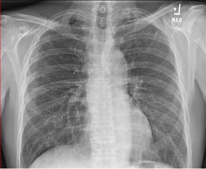
On admission she had a temperature of 39.6º C, was tachycardia and hypotensive requiring vasopressors. Labs were remarkable for a white count of 25,000 cells/mcL. Chest x-ray showed right-sided infiltrate and pleural effusion (Figure 1).

Figure 1. Chest x-ray on presentation.

Bronchoscopy and thoracentesis was performed upon admission. The pleural fluid wasexudative with a glucose of 78 and no suggestion of loculations on chest x-ray or ultrasound. The patient was started on therapy for community-acquired pneumonia.

On day 4 after admission, the patient had increasing sinus tachycardia, hypotension and was worsening despite being on antimicrobial therapy. A CT angiogram of the chest was performed (Figure 2).

Figure 2. Initial CT scan on day 4 of admission. Panel A: axial view showing pneumonia and right pleural effusion. Panel B: coronal view.

CT angiogram was negative for pulmonary embolism and a percutaneous chest tube was placed on day 4 for drainage of pleural effusion due to development of loculations. On day 7, the pleural fluid from initial thoracentesis grew acid-fast bacteria identified as Mycobacterium fortuitum.

Bronchoscopy was performed on day 8 and there was no endobronchial obstruction.

Bronchoscopic alveolar lavage cultures grew Mycobacterium fortuitum. She had no history of bronchiectasis, skin infection, or immunoglobulin deficiency. Treatment with amikacin and levofloxacin was initiated based on susceptibilities.

The pleural chest tube was removed on day 14 (Figure 3). At this time the patient was transferred to a skilled nursing facility.

Figure 3. CT scan on day 13 prior to chest tube removal. Panel A: axial view. Panel B: coronal view.

The patient continued antibiotic treatment for Mycobacterium fortuitum with amikacin and levofloxacin, however, serial sputum cultures remained positive. On day 25, in the skilled nursing facility, the patient developed respiratory failure due to increased right effusion and worsening pneumonia. She was transferred to our facility were she was intubated and a new right-sided chest tube was placed. After placement of chest tube and drainage the right lung did not expand. Decompensation was felt to be related to the inadequate evacuation of the empyema with plans to solely continue antimicrobial therapies by the outside facility.

Figure 4. CT scan on day 30 showing trapped lung. Panel A: axial view. Panel B: coronal view. 

Repeat pleural fluid cultures and BAL once again grew Mycobacterium fortuitum. She was taken for decortication and right middle and lower lobe resection by thoracic surgery. Due to extensive disease the patient required right thoracotomy, decortication, parietal pleurectomy, right middle lobectomy, and wedge resection of a right lower lobe lung abscess.

The lung pathology is shown below and was consistent with lipoid pneumonia (Figure 5).

Figure 5. Panels A & B: CD 163 stains showing lipid present within histiocytes. Panels C & D: histology demonstrating severe lipoid pneumonia. Panels E & F: Granulomatous inflammation with giant cells. Panel G: pleura. Panel H: abscess.

There were no mycobacteria cultured on the lung biopsy. There were areas of both acute and chronic fibrosis noted on pathology report along with areas of acute interstitial pneumonitis and granulomatous inflammation.

During post-operative phase the patient confirmed that she was drinking mineral oil chronically for treatment of constipation. Repeat sputum cultures 7 days post operatively were negative for Mycobacterium fortuitum. She continued to improve with treatment of Mycobacterium fortuitum and postoperative cultures remained negative. She was able to liberate from the ventilator and returned home at after a prolonged course of rehabilitation.

Lipoid Pneumonia and Associated Mycobacterial Infection

The association between acid-fast bacteria and lipoid pneumonia was first reported in 1925 and since case reports have been noted. In 1953, a case report and literature review documented six cases of “saprophytic” mycobacteria was noted in conjunction with lipoid pneumonia. It was observed at this time that the fatty environment of lipoid pneumonia might assist with the growth of mycobacterium (1). Since then, intermittent case reports have been published reporting lipoid pneumonia with atypical mycobacteria.

There are two main categories of lipoid pneumonia, endogenous and exogenous. The endogenous form is also known as cholesterol pneumonia or golden pneumonia. It is associated with lysis of lung tissue distal to obstruction due to malignancy, fat storage disease such as Neiman-Pick or Gaucher's, medications and therapies including chemotherapeutic agents, amiodarone and radiation therapy. Pulmonary alveolar proteinosis has also been reported in idiopathic cases with granulomatosis with polyangiitis and connective tissue diseases (2-4). In polarized light microscopy after staining with sulfuric and acetic acid, the sample reveals cholesterol crystals, which is diagnostic of endogenous lipoid pneumonia (3).

Exogenous lipoid pneumonia occurs when external substances enter the lungs due to inhalation or aspiration (3). Cases have been reported from mineral oil, paraffin use, oil based nasal drops, total parenteral nutrition, mineral oil nose drops, black fat tobacco smoking, milk, and liquid hydrocarbons used by flame blowers (2-6). The pulmonary reaction to each substance varies. For example, mineral oils are fairly inert and less likely to produce alveolar inflammation, where milk fats are hydrolyzed by lung lipases leading to a significant inflammatory response (2).

The clinical presentation and appearance of lipoid pneumonia is variable from consolidation to effusion to nodule. Nodules from lipids may have elevated standardized uptake value (SUV) on positron emission tomography (PET) scan. The BAL from lipoid pneumonia may demonstrate lipid laden foamy macrophages (2). Mineral oil granuloma (paraffinoma) also can present as a spiculated mass mimicking malignancy.

Mineral oil is notorious for causing lipoid pneumonia by aspiration for several reasons. First, it floats on the column of undigested material in the esophagus so it is first to be aspirated (5); secondly, it impairs phagocytosis at the alveolar level; and lastly, it inhibits the cough reflex and motor function of ciliated mucosa (7).

The impairment of phagocytosis associated with lipoid pneumonia is thought to be a contributing factor in why atypical mycobacterium strives in the lipid rich environment of lipoid pneumonia (5,6). Malnutrition is also thought to be a component of risk as it due to impairment in cell mediated immunity (6). Lipid acts as mechanical protection for the mycobacteria favoring tissue necrosis facilitating secondary infection. Also it is thought that lipids may activate the cell walls of the atypical mycobacteria leading to increased virulence of the mycolic acids within the wall of the bacteria (8).

Mycobacterium fortuitum rarely causes pulmonary disease unless associated with lipoid pneumonia. This is often related to gastroesophageal disease and chronic vomiting and aspiration of contents. It is typically associated with skin and soft tissue infections and is a rapid growing mycobacterium and most frequently found in water and soil (2,8,9)

This case demonstrates an atypical presentation of lipoid pneumonia and Mycobacterium fortuitum infection leading to septic shock and ventilator failure. Although the association of lipoid pneumonia and mycobacterial infections is well documented, the rapid and acute decline in this patient’s clinical status is unusual. This can be attributed to incomplete drainage of the initial empyema prior to transfer to the skilled nursing facility.

The etiology of the lipoid pneumonia was chronic aspiration of mineral oil producing an ideal environment for growth of Mycobacterium fortuitum. The absence of bronchiectasis, immunoglobin deficiency, skin infections should prompt further evaluation for abnormal lung architecture serving as a nidus for Mycobacterium fortuitum Infection. In our case, failure to improve is attributed to a persistent nidus for infection. We advocate resection of diseased lung segments of lipoid pneumonia to facilitate successful treatment of Mycobacterium fortuitum. In conclusion, if a patient has lipoid pneumonia with signs of clinical infection, the possibility of rapidly growing mycobacterium such as M. fortuitum should be considered.

References

  1. Gibson JB. Infection of the lungs by saprophytic mycobacteria in achalasia of the cardia, with report of a fatal case showing lipoid pneumonia due to milk. J Pathol Bacteriol. 1953;65(1):239-51. [CrossRef] [PubMed]
  2. Hasan A, Swamy T. Nocardia and Mycobacterium fortuitum infection in a case of lipoid pneumonia. Respiratory Medicine CME 2011: 75-78. [CrossRef]
  3. Betancourt SL, Martinez-Jimenez S, Rossi SE, Truong MT, Carrillo J, Erasmus JJ. Lipoid pneumonia: spectrum of clinical and radiologic manifestations. AJR Am J Roentgenol. 2010;194(1):103-9. [CrossRef] [PubMed]
  4. Harris K, Chalhoub M, Maroun R, Abi-Fadel F, Zhao F. Lipoid pneumonia: a challenging diagnosis. Heart Lung. 2011;40(6):580-4. [CrossRef] [PubMed]
  5. Hughes RL, Freilich RA, Bytell DE, Craig RM, Moran JM. Clinical conference in pulmonary disease. Aspiration and occult esophageal disorders. Chest. 1981;80(4):489-95. [CrossRef] [PubMed]
  6. Tranovich VL, Buesching WJ, Becker WJ. Pathologic quiz case. Chronic pneumonia after gastrectomy. Pathologic diagnosis: chronic aspiration lipoid pneumonia with Mycobacterium abscessus. Arch Pathol Lab Med. 2001;125(7):976-8. [PubMed]
  7. Jouannic I, Desrues B, Léna H, Quinquenel ML, Donnio PY, Delaval P. Exogenous lipoid pneumonia complicated by Mycobacterium fortuitum and Aspergillus fumigatus infections. Eur Respir J. 1996;9(1):172-4. [Pubmed]
  8. Couto SS, Artacho CA. Mycobacterium fortuitum pneumonia in a cat and the role of lipid in the pathogenesis of atypical mycobacterial infections. Vet Pathol. 2007;44(4):543-6. [CrossRef] [PubMed]
  9. Vadakekalam J, Ward MJ. Mycobacterium fortuitum lung abscess treated with ciprofloxacin. Thorax. 1991;46(10):737-8. [CrossRef] [PubMed] 

Cite as: Till S, Mathew M, Liao D-W, Ramirez C. Why chronic constipation may be harmful to your lungs: a case report and review of lipoid pneumonia and mycobacterium fortuitum leading to acute respiratory failure and septic shock. Southwest J Pulm Crit Care. 2015;11(4):193-9. doi: http://dx.doi.org/10.13175/swjpcc118-15 PDF 

Thursday
Oct152015

Traumatic Hemoptysis Complicating Pulmonary Amyloidosis

Erwan Oehler, MD1

Charlotte Courtois, MD2 

Florent Valour, MD1


1Department of Internal Medicine

2Department of Pulmonary Medicine

French Polynesia Hospital Center

98716 Pirae, Tahiti

French Polynesia


Case Presentation

We present a 74-year-old man admitted to hospital for a fall occurring at home. His past medical history included histologically-proven pulmonary amyloidosis followed for fifteen years (Figure 1A), without involvement of other organs.

Figure 1A. Frontal chest radiography shows bilateral confluent, somewhat nodular and dense-appearing opacities with a background of faint linear and reticular opacities.

At admission, he complained of left chest pain related to a rib fracture (Figure 1B, arrow).

Figure 1B. Detail radiograph of the left upper thorax shows a fracture (arrow) of a posterolateral rib, superimposed on the background of dense-appearing linear and nodular parenchymal disease.

The next day, he presented with moderate hemoptysis, prompting performance of thoracic CT (Figure 1C and D) which showed a cavity filled with material of soft tissue attenuation.

Figure 1C and D. Axial thoracic CT displayed in soft tissue windows shows extensive bilateral nodular hyperattenuating tissue consistent with alveolar septal / diffuse pulmonary parenchymal amyloidosis. A cystic lesion with internal, dependent soft tissue attenuation (arrow, D) is present, consistent with a hematoma.

This soft tissue-filled cavity was located at the same level as the rib fracture, surrounded by calcified tissue, and presumably reflected a pulmonary parenchymal hematoma resulting from traumatically induced laceration of the inelastic calcified lung tissue.

Discussion

Pulmonary amyloidosis is a rare disease resulting from the extracellular deposition of insoluble fibrillar proteins aggregating in a β–pleated sheet configuration (1). Amyloidosis is classified according to the chemistry of the amyloid protein as AA secondary amyloidosis (SAA protein) -often related to chronic inflammatory disease- AL amyloidosis (monoclonal immunoglobulin light chains of the lambda or kappa type)-secondary to B lymphoproliferative disorders-and hereditary or familial amyloidosis (transthyretin and gelsolin). Dialysis-associated amyloidosis (βR2R microglobulinemia) and “senile” amyloidosis SAA (wild-type transthyretin) are also recognized. Pulmonary amyloidosis may occur in three forms: tracheobronchial, nodular parenchymal and alveolar septal / diffuse parenchymal patterns (2). The two first forms (which include primitive pulmonary amyloidosis) are often remain localized to the respiratory system, whereas the alveolar septal / diffuse parenchymal form of amyloidosis, whose prognosis is more severe, often presents in a systemically. Parenchymal amyloid nodules grow slowly and generally remain asymptomatic but patients may also present with dyspnea, cough, hemoptysis or recurrent pneumonia (3).

References

  1. Chu H, Zhao L, Zhang Z, Gui T, Yi X, Sun X. Clinical characteristics of amyloidosis with isolated respiratory system involvement: A review of 13 cases. Ann Thorac Med. 2012 (4):243-9. [CrossRef] [Pubmed]
  2. Gilmore JD, Hawkins PN. Amyloidosis and the respiratory tract. Thorax. 1999;54:444-51. [CrossRef] [PubMed]
  3. Vieira IG, Marchiori E, Zanetti G, Cabral RF, Takayassu TC, Spilberg G, Batista RR. Pulmonary amyloidosis with calcified nodules and masses - a six-year computed tomography follow-up: a case report. Cases J. 2009;2:6540. [CrossRef] [PubMed]

Cite as: Oehler E, Courtois C, Valour F. Traumatic hemoptysis complicating pulmonary amyloidosis. Southwest J Pulm Crit Care. 2015;11(4):173-5. doi: http://dx.doi.org/10.13175/swjpcc133-15 PDF

Tuesday
Oct132015

Staphylococcus aureus Sternal Osteomyelitis: a Rare Cause of Chest Pain

Manjinder Kaur DO

Courtney Walker DO

Emily S. Nia MD

Jeffrey R. Lisse MD


Department of Medicine

Banner University Medical Center

Tucson, AZ USA


Abstract

Chest pain is a common presenting symptom with a broad differential. Life-threatening cardiac and pulmonary etiologies of chest pain should be evaluated first. However, it is critical to perform a thorough assessment for other sources of chest pain in order to limit morbidity and mortality from less common causes. We present a rare case of a previously healthy 45 year old man who presented with focal, substernal, reproducible chest pain and Staphylococcus aureus bacteremia who was later found to have primary Staphylococcus aureus sternal osteomyelitis.

Case Report

A 45 year old previously healthy man presented to the emergency department with sudden onset substernal chest pain of two days duration. The pain was described as constant, achy, worsened with movement, and improved with lying still.  Palpation of the manubrium reproduced pain and was associated with an appreciable “bump”. The patient denied recent trauma or surgery and reported no fevers, weight loss, night sweats, cough, or history of intravenous drug use. He had multiple tattoos covering his thorax and abdomen obtained while incarcerated twenty years prior to admission. On examination, the patient was uncomfortable due to severe sternal pain. He was diaphoretic, tachycardic, tachypneic, and afebrile. His manubrium was tender to palpation and the overlying skin was warm and mildly swollen without apparent erythema, induration, or drainage. Laboratory results were remarkable for leukocytosis of 18,4000/uL with 92% neutrophils, serial troponins less than 0.01 ng/mL, ESR 15 mm/hr, c-reactive protein (CRP) 13.40 mg/dL, nonreactive HIV antibodies, and positive hepatitis C virus (HCV) antibody with detectable but unquantifiable HCV RNA. Electrocardiogram showed normal sinus rhythm without ischemia. Bibasilar atelectasis was appreciated on chest x-ray and chest CT with contrast revealed no bone or chest wall lesions. Sternum MR with contrast (Figure 1) showed enhancing edema in the subcutaneous soft tissues overlying the sternomanubrial joint with extension into the pectoralis major musculature symmetrically without abscess or bony involvement.

Figure 1. Sagittal and axial T2 fat sat images (A and B) demonstrate inflammatory changes involving the soft tissues overlying the sternum including the pectoralis muscles bilaterally. Sagittal and axial T1 post contrast images (C and D) demonstrate avid enhancement involving the soft tissues overlying the sternum consistent with phlegmonous change without a rim enhancing loculated fluid collection to suggest an abscess formation. No underlying osseous involvement is present. A tissue marker corresponds to the patient’s site of pain. 

On day two of admission, blood culture results were reported positive for Staphylococcus aureus oxacillin susceptible (MSSA). Positive blood cultures persisted despite appropriate antibiotics. A transesophageal echocardiogram (TEE) was performed and showed no vegetations. Although chest imaging was negative for osteomyelitis, the persistent bacteremia and focal sternomanubrial pain was clinically suggestive of primary sternal osteomyelitis. The patient was discharged to home and completed a six week course of intravenous cefazolin for presumed MSSA sternal osteomyelitis.  

Repeat MR sternum performed eight weeks after initial presentation showed osteomyelitis across the sternomanubrial joint with improved soft tissue edema
(Figure 2).


Figure 2. Sagittal and axial T2 fat sat images (A and B) demonstrate interval improvement in inflammatory changes involving the soft tissues overlying the sternum with persistent edema present at the sternomanubrial joint (red arrow). Sagittal T1 image (C) demonstrates focal hypointense bone marrow about the sternomanubrial joint (red arrow). Sagittal and axial T1 post contrast images (D and E) demonstrate enhancement of the sternomanubrial joint (red arrow). Overall findings are consistent with osteomyelitis of the sternomanubrial joint.

Given that the patient had completed six weeks of parenteral antibiotic therapy, his sternal chest pain had resolved, and CRP had normalized, additional antibiotics were not prescribed and the patient was asked to follow up with his primary care provider as needed. There was no incidence of further complication and the patient was diagnosed with primary MSSA sternal osteomyelitis.

Discussion       

Primary osteomyelitis of the sternum in immunocompetent patients is extremely rare, accounting for 0.3% of all cases of osteomyelitis reported in the literature (1). Common risk factors for primary sternal osteomyelitis are trauma, pneumonia, diabetes, immunodeficiency, or history of IV drug use (2,3). Our patient had none of these risk factors. Risks for secondary sternal osteomyelitis are due to complications from sternal incision post-thoracic surgery(1-3). Staphylococcus aureus is the most common organism of both primary and secondary sternal osteomyelitis (2).

Early diagnosis of acute osteomyelitis is critical in order to prevent necrosis of bone, as well as other local and systemic complications, from delayed antibiotic therapy. Multiple imaging modalities are available to confirm the presumed clinical diagnosis of osteomyelitis. MRI is 82% to 100% sensitive and 75% to 96% specific and is considered the gold standard in diagnosis of acute osteomyelitis (4). However, as evidenced by our case, imaging findings may lag behind clinical presentation. Clinicians need to consider primary osteomyelitis in the differential diagnosis of a young patient who presents with focal sternal chest pain, swelling, and bacteremia. A strong index of suspicion for acute osteomyelitis is needed in order to promptly initiate antibiotic therapy to reduce morbidity and mortality associated with untreated osteomyelitis (1,2).

References

  1. de Nadai TR, Daniel RF, de Nadai MN, da Rocha JJ, Féres O. Hyperbaric oxygen therapy for primary sternal osteomyelitis: a case report. J Med Case Rep. 2013;7:167. [CrossRef] [PubMed]
  2. Gill EA Jr, Stevens DL. Primary sternal osteomyelitis. West J Med. 1989;151(2):199-203. [PubMed]
  3. Vacek TP, Rehman S, Yu S, Moza A, Assaly R. Another cause of chest pain: Staphylococcus aureus sternal osteomyelitis in an otherwise healthy adult. Int Med Case Rep J. 2014;7:133-7. [CrossRef] [PubMed]
  4. Pineda C, Espinosa R, Pena A. Radiographic imaging in osteomyelitis: the role of plain radiography, computed tomography, ultrasonography, magnetic resonance imaging, and scintigraphy. Semin Plast Surg. 2009;23(2):80-9. [CrossRef] [PubMed] 

Cite as: Kaur M, Walker C, Nia ES, Lisse JR. Staphylococcus aureus sternal osteomyelitis: a rare cause of chest pain. Southwest J Pulm Crit Care. 2015;11(4):167-70. doi: http://dx.doi.org/10.13175/swjpcc131-15 PDF  

Saturday
Oct102015

Safety and Complications of Bronchoscopy in an Adult Intensive Care Unit

Aarthi Ganesh, MBBS1

Nirmal Singh, MBBS, MPH2

Gordon E. Carr, MD1


1Department of Pulmonary & Critical Care

2Department of Internal Medicine

University of Arizona

Tucson, Arizona


Abstract

Background: Bronchoscopy is a common procedure performed in adult intensive care units (ICU). However, very few studies report the safety and complications of the bronchoscopy and related procedures performed on critically ill patients. The primary aim of this study was to determine the incidence of complications following ICU bronchoscopy.

Methods: We conducted a retrospective chart review of patients admitted to an adult ICU and underwent bronchoscopy with or without bronchoalveolar lavage (BAL) and other bronchoscopic procedures. Data included patient demographics, APACHE II score, hemodynamics, comorbidities, type of ventilation and procedure performed. Data from BAL, including cellular differential and microbiology, were also collected.

Results: We identified 120 patient charts between November 2011 to March 2012. The most common procedure was bronchoscopy with BAL (62%) to evaluate for pneumonia (58%). Other procedures included transbronchial biopsy, APC and cryotherapy, balloon and stent placement, endobronchial biopsy and EBUS. Complications occurred in 18% of the patients, with hypoxia being the most common (7.5%). No deaths occurred related to the procedures. Nine percent of patients who had BAL or inspection had complications compared to 29% who underwent other procedures. Subgroup analysis conducted on patients undergoing BAL revealed significantly higher neutrophil counts (p=0.001) and higher APACHE II score (p=0.02) among those with BAL positive for bacteria and co-infection.

Conclusion: Bronchoscopy with BAL and inspection is relatively safe procedure even in critically ill patients. However, other interventional bronchoscopic procedures should be performed with caution in the ICU.

Abbreviations:

ICU: Intensive care unit

BAL: Bronchoalveolar lavage

EBUS: Endobronchial Ultrasound

APC: Argon Plasma Coagulation

SBP: Systolic Blood Pressure

CI: Confidence Interval

IP: Interventional pulmonary

MAP: Mean arterial pressure

SD: Standard deviation

CHF: Congestive heart Failure

COPD: Chronic Obstructive Pulmonary Disease

ILD: Interstitial Lung Disease

ET: Endotracheal

Introduction

Fiberoptic bronchoscopy is a commonly performed procedure in the medical intensive care unit (ICU). Prior studies have indicated that bronchoscopy is generally safe, making it a relatively low-risk procedure in appropriately selected ICU patients (1-3). Most prior studies reporting the safety of bronchoscopy were performed in early 1990s. The rates of complications or adverse events in these earlier studies ranged from 2% to 40% (2,4-6). The primary aim of this study was to assess the incidence of complications in ICU patients undergoing bronchoscopy in the contemporary era.

Methods

The study was approved by the Institutional Review Board at the University of Arizona. We conducted a retrospective chart review of patients, 18 years or older, admitted to the adult medical intensive care unit, who underwent bronchoscopy with or without bronchoalveolar lavage (BAL) and other bronchoscopic procedures from November 1, 2011 to March 31, 2012. The other bronchoscopic procedures included transbronchial biopsies, endobronchial ultrasound (EBUS) guided biopsy, argon plasma coagulation (APC) and cryotherapy, balloon dilatation with stenting, and endobronchial biopsy. We excluded patients with incomplete charts, and patients who had bronchoscopy as a part of percutaneous tracheostomy procedure. Data included patient demographics, APACHE II scores, hemodynamics, co-morbidities, type of ventilation, type of procedure performed and the complications. Sedation used in the procedures included propofol or midazolam with fentanyl for analgesia. BAL results, including cellular differential and microbiology studies, were also collected. We used pre-specified definitions to assess for complications. We defined hypotension as reduction in systolic blood pressure (SBP) by >20 mm Hg or when a patient required vasopressors to maintain a mean arterial pressure (MAP) > 60 mm Hg during or after the procedure. Hypoxia was defined by drop in saturation to < 90% or when the FiO2 requirement increased by > 20% for more than 2 hours after the procedure. Hemorrhage was indicated as per the procedure note by the bronchoscopist or when the note indicated use of epinephrine or when additional procedures needed to be performed to control the bleeding. During the procedure all the patients FiO2 was increased but was turned down to their previous ventilatory settings unless there was significant hypoxia.

Statistical analysis was performed using STATA/IC 13.1 (StataCorp LP, Texas). Numerical variables are expressed as mean ± standard deviation (SD). Ninety-five percent confidence intervals (CIs) were calculated where appropriate. Univariate comparisons between patients who did and did not develop complications were calculated using a χ2 test or Fischer's exact test for categorical variables and a 2-sample t test for continuous variables applying central limit theorem. All statistical testing was two-tailed with significance level set at the alpha level of ≤0.05.

Results

We identified 140 patients who underwent ICU bronchoscopy during the study period. Eighteen patients were excluded due to incomplete information. Two charts were excluded as the bronchoscopy was performed for percutaneous tracheostomy. Table 1 shows the baseline characteristics of patients undergoing ICU bronchoscopy.

Table 1. Baseline Characteristics of Patients Prior to Bronchoscopy

Key: CAD: Coronary Artery Disease

        CHF: Congestive Heart Failure

        COPD: Chronic obstructive pulmonary disease

        FiO2: Oxygen required

        ILD: Interstitial Lung Disease

        MAP: Mean arterial pressure

        NM Disease: Neuromuscular disease

Sixty-nine percent of the patients were male and average age was 52 ± 16 years. The average APACHE II score was 18 ± 6 with a median of 18 and 88% of the patients were intubated and mechanically ventilated. The mean percentage oxygen (FiO2) requirement in the patients prior to the procedure was 63% ± 26. Sixty-three percent of the patients were immunocompromised, likely related to the large proportion of lung transplant recipients in our study population. Fifty-four percent also had chronic lung disease including chronic obstructive pulmonary disease (COPD) and interstitial lung disease (ILD). Other common co-morbidities included cardiovascular disease including congestive heart failure (CHF) and arrhythmias, malignancy and neuromuscular diseases. Table II shows the indications for ICU bronchoscopy. The most common indication for the procedure was to evaluate for pneumonia or infiltrate in 87 cases (72%), followed by atelectasis/ collapse/ secretions in 19 cases (15.8%) (Table 2).

Table 2. Indications For Procedures

Other indications included tracheal or airway diseases, which included tracheal stenosis, upper airway obstruction, tracheal mass and bronchopleural fistula in 11 (8%) and hemoptysis (2%). The most common procedures performed were bronchoscopy with BAL in 75 (62%) and inspection in 31 (26%) (Table 3).

Table 3. Procedures

Key:  APC: Argon plasma coagulation

         BAL: Bronchoalveolar lavage

         Cryo: Cryotherapy

         EBUS: Endobronchial ultrasound

         ET: Endotracheal tube

Other procedures included transbronchial biopsy, APC and cryotherapy, balloon and stent placement, endobronchial biopsy and EBUS.

Table 4 shows the complications resulting from ICU bronchoscopy in this study population.

Table 4. Complications

Twenty two complications occurred during or within 2 hours after the procedure (18%), with hypoxia being the most common (7.5%). Hypoxia in two patients occurred secondary to hemorrhage. Pneumothorax was seen in one patient who underwent transbronchial biopsy with no fluoroscopic guidance. Hypotension which needed treatment with fluids or vasopressors occurred in 5.8% and hemorrhage in 3.3%. Hemorrhage was unrelated to coagulopathy in the patients. Significant bradycardia requiring treatment with atropine occurred in one patient. No deaths were reported related to the procedures. None of the procedures had to be terminated secondary to the complications. More adverse events were seen among the patients who underwent other bronchoscopic procedures (29%) than those undergoing BAL or inspection only (9%), though this was not statistically significant (p = 0.07).

As depicted in Table 5, none of the complications were significantly affected by the underlying comorbidities or the APACHE scores.

Table 5. Patient Characteristics Stratified by Complications

Key: BAL: Bronchoalveolar lavage

       MAP: Mean Arterial Pressure

Complications were not significantly associated with the amount of oxygen required (FiO2) and the mode of ventilation which the patients were on prior to the procedure. Similarly, neither the mean arterial pressure before the procedure or coagulopathy influenced the rate of complications. Hospital mortality was not different in the group with or without complications.

Figure 1 and Table 6 show the BAL cell differential.

Figure 1. BAL differential in culture with normal respiratory flora (0), bacteria (1), Viral (2), Fungal (3) and Co-infection (4). Each bar represents the differential in percentage.

Key: BAL: Bronchoalveolar lavage

          BAL N: Neutrophil count in BAL (in percentage)

          BAL L: Lymphocyte count in BAL (in percentage)

          BAL M: Macrophages count in BAL (in percentage)

          BAL E: Eosinophils count in BAL (in percentage)

Table 6. Bronchoalveolar Lavage Differential

Patients found to have bacterial pneumonia or mixed viral and bacterial infection had significantly higher neutrophil counts (mean BAL neutrophil count 82% for bacterial infection, and 80% for co-infections) than other patients (p=0.001) (Figure 2).

Figure 2. Neutrophil predominance in bacterial pneumonia. KEY: BAL-N: Bronchoalveolar lavage, neutrophil differential (in percentage).

These patients also had a higher APACHE II score (p=0.02). Hospital mortality was higher among those with BAL positive for bacteria (p= 0.012). Mortality was also significantly higher among patients with underlying malignancy (p= 0.002).

Discussion

In our study of 120 ICU bronchoscopies, we found a complication rate of 18%. No deaths were observed in this study. Hypoxia was the most common adverse event in our study, occurring in 9 procedures (7.5%) as has been noticed in the previous studies. Introduction of a bronchoscope through an endotracheal (ET) tube is known to cause airway obstruction resulting in increasing intra-tracheal pressures and variation in respiratory physiology (6). Almost all the patients who were mechanically ventilated had a size 7.5 - 8.5 ET tube or had tracheostomy in place. As in prior studies, BAL performed for evaluation of pneumonia and atelectasis were the two most common indications of the procedure (72% and 15.8% respectively) in our study (1-7). Even though bronchoscopy has not shown to be routinely superior to chest physiotherapy, certain subset of patient population may benefit from it (3,8,9). Improvement in oxygenation has been shown to occur in certain earlier studies (10,11).

Hypotension is also a known complication occurring during bronchoscopy. Our study had 7 events (5.8%) of hypotension needing vasopressor or fluid infusion. This was likely related to the sedation. Hypertension was observed in one case and bradycardia requiring treatment was seen in one. Cardiovascular abnormalities associated with bronchoscopy is generally related to the sympathetic surge happening during the procedure and the hypoxia (12-14). Per earlier studies, the complication rate of transbronchial biopsies in mechanically ventilated patients range between 0-15% (15,16,17). But it is relatively safe in comparison to open lung biopsy.

With the advent of newer technology, there has been an increase in the number of other bronchoscopic interventional pulmonary (IP) procedures, including endobronchial ablative therapies such as APC and cryotherapy. Endobronchial lesions occupying more than 50% of the airway lumen can alter the airway physiology and result in hypoxia, ventilation perfusion mismatch and hence respiratory failure. Use of ablative therapies can potentially reverse this (18). APC has been an useful tool to remove endobronchial lesions and relieve obstruction. It has been shown to be efficient and relatively safe in outpatient setting, but APC on mechanically ventilated patients has not been very well studied (19). APC in mechanically ventilated patient requires decrease in the FiO2 to less than or equal to 40%. Complications related to IP procedures performed specifically in patients requiring mechanical ventilation are difficult to assess  from the available literature (20). However, given the complexity of these cases and underlying illness, usually the complications are minor. In our study, interventional bronchoscopy procedures like APC, cryotherapy was to relieve airway obstruction which was the cause of mechanical ventilation. In our study, APC case was associated with hemorrhage. The balloon dilatation and stenting which was performed for a case of tracheal stenosis arising from malignancy. This was not associated with any complications related to the procedure in our study. Further study is needed to refine our understanding of the risks of advanced bronchoscopic techniques in ICU patients.

Procedures like EBUS are usually not done in critically ill patients. There are no studies which have looked into the use of and complications of performing EBUS in critically ill patients. Bhaskar et al. (21) report the use of esophageal access for mediastinal sampling through EBUS in ICU patients for the reason of causing hypoxia and changes in airway physiology with the EBUS scope in airway. Our study had one patient who had an EBUS for lung mass and this was not associated with any complications.

Subgroup analysis in our study showed the presence of neutrophilic predominance with neutrophil count of >80% in the BAL differential in patients diagnosed with bacterial infections and co-infections compared to those with viral/ fungal or mixed flora (p=0.001). This was similar to results from earlier studies (22,23). Neutrophilic pleocytosis in BAL fluid is frequently found in patients with pneumonia. As the neutrophil count is higher in bacterial pneumonia, it can indicate towards a differential of bacterial pneumonia even prior to the final microbiology results. Hence BAL differential may be complimentary to final culture results and maybe helpful to initiate or discontinue antibiotics in critically ill patients. Mortality among critically ill patients with bacterial pneumonia was higher compared to others (p=0.012). These patients tend to be sicker with higher APACHE II scores.

The weaknesses of the study includes the fact that it was retrospective chart review. The total number is small, and the number of the IP procedures performed is even smaller. Hence it is important that more studies should be conducted looking into the safety and complications of IP procedures in critically ill patients.

Conclusion

Our study looked into the fiberoptic bronchoscopy with BAL and inspection as well as other therapeutic procedures done in the critically ill patients. It indicates that even in critically ill patients, bronchoscopy with inspection and BAL is safe. Other interventional pulmonary procedures may have more complications. Even though the number of IP procedures performed in the study is low, the evidence of slightly more number of complications with these procedures indicates the need for caution before attempting them in the critically ill patients.

References

  1. Barrett CR Jr. Flexible fiberoptic bronchoscopy in the critically ill patient. Methodology and indications. Chest. 1978;73(5 Suppl):746-9. [CrossRef] [PubMed]
  2. Hertz MI, Woodward ME, Gross CR, Swart M, Marcy TW, Bitterman PB. Safety of bronchoalveolar lavage in the critically ill, mechanically ventilated patient. Crit Care Med. 1991;19(12):1526-32. [CrossRef] [PubMed]
  3. Olopade CO, Prakash UB. Bronchoscopy in the critical-care unit. Mayo Clin Proc. 1989;64(10):1255-63. [CrossRef] [PubMed]
  4. Steinberg KP, Mitchell DR, Maunder RJ, Milberg JA, Whitcomb ME, Hudson LD. Safety of bronchoalveolar lavage in patients with adult respiratory distress syndrome. Am Rev Respir Dis. 1993;148(3):556-61. [CrossRef] [PubMed]
  5. Prebil SE, Andrews J, Cribbs SK, Martin GS, Esper A. Safety of research bronchoscopy in critically ill patients. J Crit Care. 2014;29(6):961-4. [CrossRef] [PubMed]
  6. Guerreiro da Cunha Fragoso E, Gonçalves JM. Role of fiberoptic bronchoscopy in intensive care unit: current practice. J Bronchology Interv Pulmonol. 2011;18(1):69-83. [CrossRef] [PubMed]
  7. Raoof S, Mehrishi S, Prakash UB. Role of bronchoscopy in modern medical intensive care unit. Clin Chest Med. 2001;22(2):241-61, vii. [CrossRef] [PubMed]
  8. Kreider ME, Lipson DA. Bronchoscopy for atelectasis in the ICU: a case report and review of the literature. Chest. 2003;124(1):344-50. [CrossRef] [PubMed]
  9. Marini JJ, Pierson DJ, Hudson LD. Acute lobar atelectasis: a prospective comparison of fiberoptic bronchoscopy and respiratory therapy. Am Rev Respir Dis. 1979;119(6):971-8. [PubMed]
  10. Snow N, Lucas AE. Bronchoscopy in the critically ill surgical patient. Am Surg. 1984;50(8):441-5. [PubMed]
  11. Stevens RP, Lillington GA, Parsons GH. Fiberoptic bronchoscopy in the intensive care unit. Heart Lung. 1981;10(6):1037-45. [PubMed]
  12. Katz AS, Michelson EL, Stawicki J, Holford FD. Cardiac arrhythmias. Frequency during fiberoptic bronchoscopy and correlation with hypoxemia. Arch Intern Med. 1981;141(5):603-6. [CrossRef] [PubMed]
  13. Lindholm CE, Ollman B, Snyder JV, Millen EG, Grenvik A. Cardiorespiratory effects of flexible fiberoptic bronchoscopy in critically ill patients. Chest. 1978;74(4):362-8. [CrossRef] [PubMed]
  14. Trouillet JL, Guiguet M, Gibert C, Fagon JY, Dreyfuss D, Blanchet F, Chastre J. Fiberoptic bronchoscopy in ventilated patients. Evaluation of cardiopulmonary risk under midazolam sedation. Chest. 1990;97(4):927-33. [CrossRef] [PubMed]
  15. Bulpa PA, Dive AM, Mertens L, Delos MA, Jamart J, Evrard PA, Gonzalez MR, Installé EJ. Combined bronchoalveolar lavage and transbronchial lung biopsy: safety and yield in ventilated patients. Eur Respir J. 2003;21(3):489-94. [CrossRef] [PubMed]
  16. O'Brien JD, Ettinger NA, Shevlin D, Kollef MH. Safety and yield of transbronchial biopsy in mechanically ventilated patients. Crit Care Med. 1997;25(3):440-6. [CrossRef] [PubMed]
  17. Casal RF, Ost DE, Eapen GA. Flexible bronchoscopy. Clin Chest Med. 2013;34(3):341-52. [CrossRef] [PubMed]
  18. Seaman JC, Musani AI. Endobronchial ablative therapies. Clin Chest Med. 2013;34(3):417-25. [CrossRef] [PubMed]
  19. Morice RC, Ece T, Ece F, Keus L. Endobronchial argon plasma coagulation for treatment of hemoptysis and neoplastic airway obstruction. Chest. 2001;119(3):781-7. [CrossRef] [PubMed]
  20. Boyd M, Rubio E. The utility of interventional pulmonary procedures in liberating patients with malignancy-associated central airway obstruction from mechanical ventilation. Lung. 2012;190(5):471-6. [CrossRef] [PubMed]
  21. Bhaskar N, Shweihat YR, Bartter T. The intubated patient with mediastinal disease--a role for esophageal access using the endobronchial ultrasound bronchoscope. J Intensive Care Med. 2014;29(1):43-6. [CrossRef] [PubMed]
  22. Stolz D, Stulz A, Müller B, Gratwohl A, Tamm M. BAL neutrophils, serum procalcitonin, and C-reactive protein to predict bacterial infection in the immunocompromised host. Chest. 2007;132(2):504-14. [CrossRef] [PubMed]
  23. Choi SH, Hong SB, Hong HL, Kim SH, Huh JW, Sung H, Lee SO, Kim MN, Jeong JY, Lim CM, Kim YS, Woo JH, Koh Y. Usefulness of cellular analysis of bronchoalveolar lavage fluid for predicting the etiology of pneumonia in critically ill patients. PLoS One. 2014;9(5):e97346. [CrossRef] [PubMed]

Cite as: Ganesh A, Singh N, Carr GE. Safety and complications of bronchoscopy in an adult intensive care unit. Southwest J Pulm Crit Care. 2015;11(4):156-66. doi: http://dx.doi.org/10.13175/swjpcc106-15 PDF

Thursday
Oct012015

October 2015 Pulmonary Case of the Month: I've Heard of Katy Perry

Kathryn E. Williams, MB

Maxwell L. Smith, MD

Philip J. Lyng, MD

Laszlo T. Vaszar, MD


Department of Pulmonary Medicine

Mayo Clinic Arizona

Scottsdale, AZ


History of Present Illness

A 45-year-old man with a history of dyslipidemia and a family history of early coronary artery disease (CAD) underwent coronary artery calcium scoring CT. He was a non-smoker and asymptomatic.

Past Medical History

In addition to his hyperlipidemia he has a history of obesity and impaired fasting glucose.

Physical Examination

His physical examination was unremarkable.

Radiography

The thoracic CT was interpreted as a low risk for CAD but there were incidental findings (Figure 1).

Figure 1. Panels A-C: Representative views from the thoracic CT scan in lung windows. Lower panel: video of thoracic CT in lung windows.

What incidental finding is not shown on thoracic CT scan. (Click on the correct answer to proceed to the second of six panels).

  1. Honeycombing
  2. Multiple small pulmonary nodules
  3. Patchy ground glass opacities
  4. Slightly enlarged mediastinal lymph nodes

Cite as: Williams KE, Smith ML, Lyng PJ, Vaszar LT. October 2015 pulmonary case of the month: I've heard of Katy Perry. Southwest J Pulm Crit Care. 2015;11(4):126-35. doi: http://dx.doi.org/10.13175/swjpcc123-15 PDF