
Correct!
5. All the above
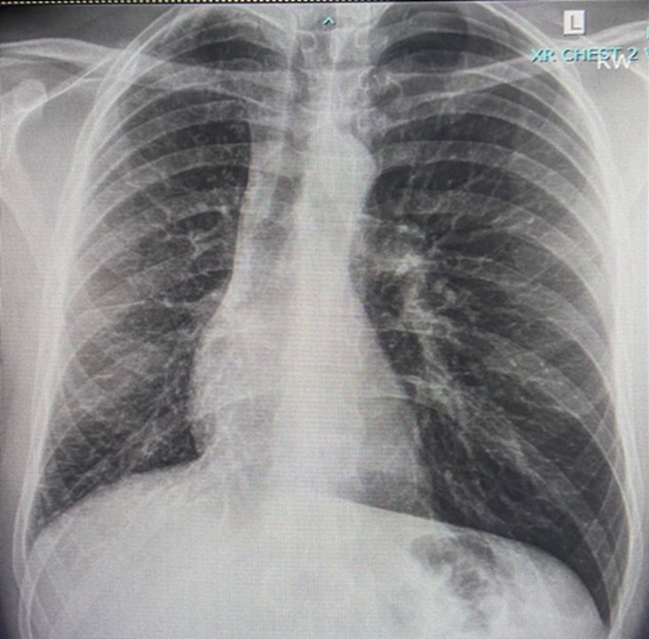
The chest radiograph was remarkable for a small right hemithorax, with mediastinal shift to the right, and the absence of infiltrates or other pathological findings (Figure 1) .

Figure 1. Admission chest X-ray. To view figure 1 in a separate, enlarged window click here .
A computerized tomography angiogram (CTA) chest ordered in the ER (Figure 2) showed no pulmonary embolism, but extensive right mediastinal and hilar soft tissue density with hypervascularity “suggestive of an infiltrative process.”

Figure 2. Selected images from CTA: A) right hemithorax volume loss, abnormal mediastinal and right peribronchial soft tissue, and absent pulmonary veins (arrow, A), B) interlobular septal thickening (arrow, B), C) prominent bronchial arteries (arrow, C). To view figure 2 in a separate, enlarged window click here .
Moderate narrowing of the right pulmonary artery, and enlargement of the left pulmonary artery were noted. The right superior and inferior pulmonary veins were “completely obliterated” at their expected sites of insertion into the left atrium. The right lung was small, without bronchial obstruction, with smooth interlobular septal thickening throughout, consistent with venous congestion. The right bronchial artery appeared hypertrophic.
Which of the following are considered in the differential of mediastinal mass / lymphadenopathy in an adult? (Click on the correct answer to be directed to the third of five pages)