Search Journal-type in search term and press enter
Southwest Pulmonary and Critical Care Fellowships
In Memoriam
Social Media

Pulmonary

Last 50 Pulmonary Postings

(Click on title to be directed to posting, most recent listed first)

March 2025 Pulmonary Case of the Month: Interstitial Lung Disease of
   Uncertain Cause
December 2024 Pulmonary Case of the Month: Two Birds in the Bush Is
   Better than One in the Hand
Glucagon‐like Peptide-1 Agonists and Smoking Cessation: A Brief Review
September 2024 Pulmonary Case of the Month: An Ounce of Prevention
   Caused a Pound of Disease
Yield and Complications of Endobronchial Ultrasound Using the Expect
   Endobronchial Ultrasound Needle
June 2024 Pulmonary Case of the Month: A Pneumo-Colic Association
March 2024 Pulmonary Case of the Month: A Nodule of a Different Color
December 2023 Pulmonary Case of the Month: A Budding Pneumonia
September 2023 Pulmonary Case of the Month: A Bone to Pick
A Case of Progressive Bleomycin Lung Toxicity Refractory to Steroid Therapy
June 2023 Pulmonary Case of the Month: An Invisible Disease
February 2023 Pulmonary Case of the Month: SCID-ing to a Diagnosis
December 2022 Pulmonary Case of the Month: New Therapy for Mediastinal
   Disease
Kaposi Sarcoma With Bilateral Chylothorax Responsive to Octreotide
September 2022 Pulmonary Case of the Month: A Sanguinary Case
Electrotonic-Cigarette or Vaping Product Use Associated Lung Injury:
   Diagnosis of Exclusion
June 2022 Pulmonary Case of the Month: A Hard Nut to Crack
March 2022 Pulmonary Case of the Month: A Sore Back Leading to 
   Sore Lungs
Diagnostic Challenges of Acute Eosinophilic Pneumonia Post Naltrexone
Injection Presenting During The COVID-19 Pandemic
Symptomatic Improvement in Cicatricial Pemphigoid of the Trachea
   Achieved with Laser Ablation Bronchoscopy
Payer Coverage of Valley Fever Diagnostic Tests
A Summary of Outpatient Recommendations for COVID-19 Patients
   and Providers December 9, 2021
December 2021 Pulmonary Case of the Month: Interstitial Lung
   Disease with Red Knuckles
Alveolopleural Fistula In COVID-19 Treated with Bronchoscopic 
   Occlusion with a Swan-Ganz Catheter
Repeat Episodes of Massive Hemoptysis Due to an Anomalous Origin 
   of the Right Bronchial Artery in a Patient with a History
   of Coccidioidomycosis
September 2021 Pulmonary Case of the Month: A 45-Year-Old Woman with
   Multiple Lung Cysts
A Case Series of Electronic or Vaping Induced Lung Injury
June 2021 Pulmonary Case of the Month: More Than a Frog in the Throat
March 2021 Pulmonary Case of the Month: Transfer for ECMO Evaluation
Association between Spirometric Parameters and Depressive Symptoms 
   in New Mexico Uranium Workers
A Population-Based Feasibility Study of Occupation and Thoracic
   Malignancies in New Mexico
Adjunctive Effects of Oral Steroids Along with Anti-Tuberculosis Drugs
   in the Management of Cervical Lymph Node Tuberculosis
Respiratory Papillomatosis with Small Cell Carcinoma: Case Report and
   Brief Review
December 2020 Pulmonary Case of the Month: Resurrection or 
   Medical Last Rites?
Results of the SWJPCC Telemedicine Questionnaire
September 2020 Pulmonary Case of the Month: An Apeeling Example
June 2020 Pulmonary Case of the Month: Twist and Shout
Case Report: The Importance of Screening for EVALI
March 2020 Pulmonary Case of the Month: Where You Look Is 
   Important
Brief Review of Coronavirus for Healthcare Professionals February 10, 2020
December 2019 Pulmonary Case of the Month: A 56-Year-Old
   Woman with Pneumonia
Severe Respiratory Disease Associated with Vaping: A Case Report
September 2019 Pulmonary Case of the Month: An HIV Patient with
   a Fever
Adherence to Prescribed Medication and Its Association with Quality of Life
Among COPD Patients Treated at a Tertiary Care Hospital in Puducherry
    – A Cross Sectional Study
June 2019 Pulmonary Case of the Month: Try, Try Again
Update and Arizona Thoracic Society Position Statement on Stem Cell 
   Therapy for Lung Disease
March 2019 Pulmonary Case of the Month: A 59-Year-Old Woman
   with Fatigue
Co-Infection with Nocardia and Mycobacterium Avium Complex (MAC)
   in a Patient with Acquired Immunodeficiency Syndrome 
Progressive Massive Fibrosis in Workers Outside the Coal Industry: A Case 
   Series from New Mexico
December 2018 Pulmonary Case of the Month: A Young Man with
   Multiple Lung Masses
Antibiotics as Anti-inflammatories in Pulmonary Diseases
September 2018 Pulmonary Case of the Month: Lung Cysts
Infected Chylothorax: A Case Report and Review
August 2018 Pulmonary Case of the Month
July 2018 Pulmonary Case of the Month
Phrenic Nerve Injury Post Catheter Ablation for Atrial Fibrillation
Evaluating a Scoring System for Predicting Thirty-Day Hospital 
   Readmissions for Chronic Obstructive Pulmonary Disease Exacerbation
Intralobar Bronchopulmonary Sequestration: A Case and Brief Review


For complete pulmonary listings click here.

The Southwest Journal of Pulmonary and Critical Care publishes articles broadly related to pulmonary medicine including thoracic surgery, transplantation, airways disease, pediatric pulmonology, anesthesiolgy, pharmacology, nursing  and more. Manuscripts may be either basic or clinical original investigations or review articles. Potential authors of review articles are encouraged to contact the editors before submission, however, unsolicited review articles will be considered.

-------------------------------------------------------------------------------------

Saturday
Apr212012

Pulmonary Nodules with Cutaneous Manifestations: A Case Report and Discussion

Christopher Strawter, MD

Pedro Quiroga, MD

Syed Zaidi, MD

Thomas Ardiles, MD


Maricopa Medical Center

Phoenix, Arizona


Abstract

The differential diagnosis of multiple pulmonary nodules is large and includes congenital and inherited disorders, malignancy, infectious etiologies, noninfectious granulomatous and inflammatory conditions,among many others. Diagnostic evaluation is aided by attention to extrapulmonary symptoms and features. We herein describe an unusual case of multiple pulmonary nodules attributed to cysticercosis and present a discussion of pathophysiologic changes related to medications and highlight the diagnostic value of extrapulmonary cutaneous features.

Case Report

History of Present Illness

A 31-year-old incarcerated Hispanic male presented with a nonproductive cough for several months and one episode of blood tinged sputum.  He admitted to weight loss and night sweats, headaches, and visual disturbances.  He was an immigrant from Honduras and lived in Arizona for the past 15 years.  He had chronic hepatitis C infection and was receiving treatment with pegylated interferon-alfa-2a (IFN-α) and ribavirin. His symptoms began one month after initiating antiviral therapy.

Physical Exam

On admission, his vital signs were stable.  Several mobile one cm cutaneous and subcutaneous nodules were palpable on the trunk and neck. Lymphadenopathy was not present.  The neurological exam revealed diplopia upon lateral gaze. The cardiopulmonary exam was unremarkable. 

Laboratory Data

Routine hematology work-up revealed leukopenia (2.3 x 10³/µL), neutropenia (1.4 x 10³/µL) without eosinophilia.  AST and ALT were 239 U/L and 265 U/L respectively. Quantiferon TB and coccidioidomycosis tests were negative.

Radiology

Plain radiograph of the chest was unremarkable.   Computed Tomography (CT) of the chest demonstrated multiple bilateral well defined nodules ranging in size from 7.2 mm to 11mm in diameter involving lung parenchyma and chest wall soft tissue structures (Figure 1). 
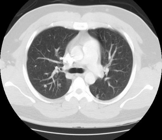
Figure 1. Computed tomography (CT) chest lung window.  Seen here are multiple, bilateral, well defined nodules involving the lung parenchyma and subcutaneous tissue.

MRI of the brain showed extensive bilateral T2 hyperintense and peripherally enhancing foci within the cerebrum, cerebellum and extraocular muscles consistent with the vesicular stage of neurocysticercosis (Figure 2). 
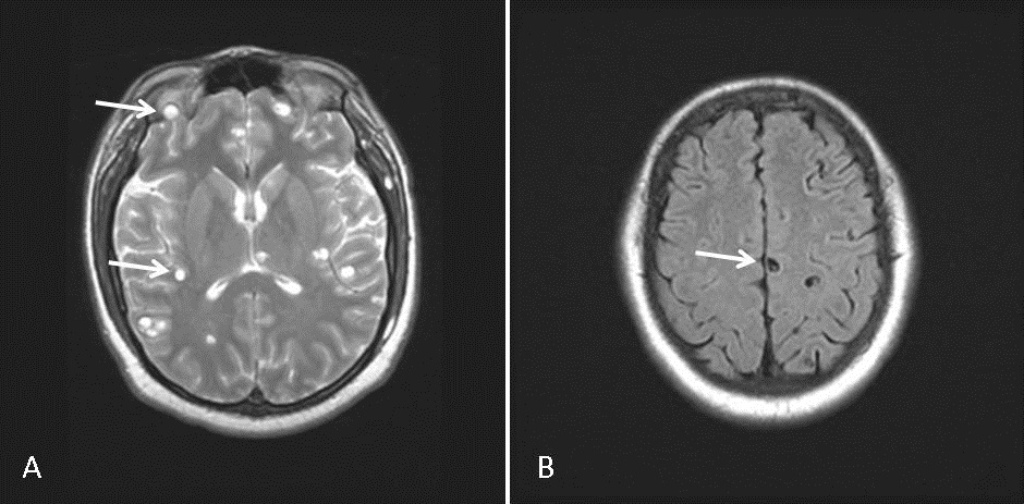
Figure 2. MRI brain.  Extensive bilateral T2 hyperintense and peripherally enhancing foci within the brain parenchyma and extraocular muscles consistent with the vesicular stage of neurocysticercosis (A). The scolex can be visualized within the cyst as a high intensity nodule giving the lesion a pathognomonic ‘hole-with-dot’ appearance (B).

Histopathology

Bronchoscopy revealed normal airways with negative bronchoalveolar lavage.  Histopathology from a needle core biopsy of a chest wall nodule revealed a cystic wall structure, consistent with cysticercosis (Figure 3).
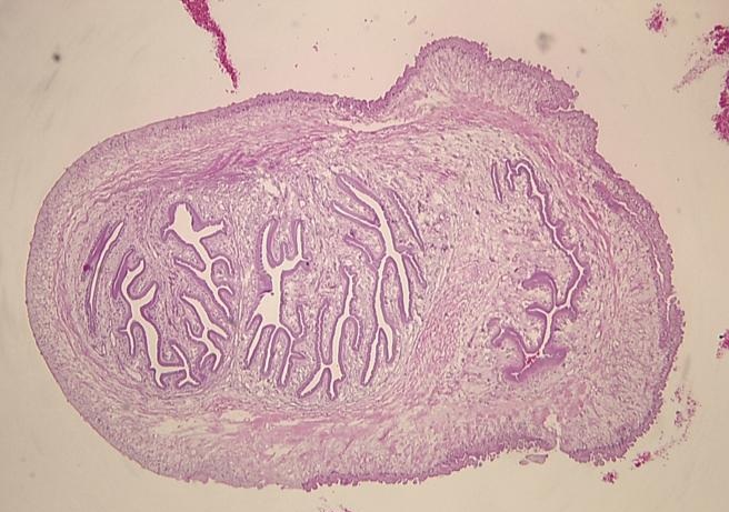
 

Figure 3. Stained microsection of a chest wall needle core biopsy showing the cystic wall structure of cysticercosis. The wall consists of 3 layers: an outer or cuticular layer, a middle cellular layer and an inner fibrillary layer.

Percutaneous lung nodule biopsy demonstrated nonspecific necrotic granulomatous tissue. 

Hospital Course

On the basis of neuroimaging and subcutaneous biopsy findings, a diagnosis of disseminated cysticercosis with pulmonary involvement was made and the patient was started on a 28-day course of albendazole therapy.  One month follow-up revealed resolution of respiratory symptoms. Repeat CT chest, 53 days post-hospitalization revealed a regression in the magnitude of the pulmonary nodules, the largest now measuring 6mm in diameter (Figure 4). 
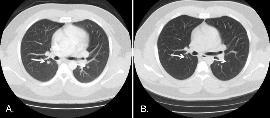
Figure 4. CT scans of the chest, lung windows. Comparing Panel A with Panel B performed 53 days later reveals a reduction in the size of the bilateral pulmonary nodules (arrows) after a 28-day course of anti-parasitic therapy with albendazole.

Regression of the pulmonary nodules was consistent with the diagnosis of pulmonary cysticercosis, in line with previous reports (1).

Discussion

The differential diagnosis of multiple pulmonary nodules is large. A variety of pulmonary disorders may affect both cutaneous and subcutaneous tissues and the lung (Table 1).
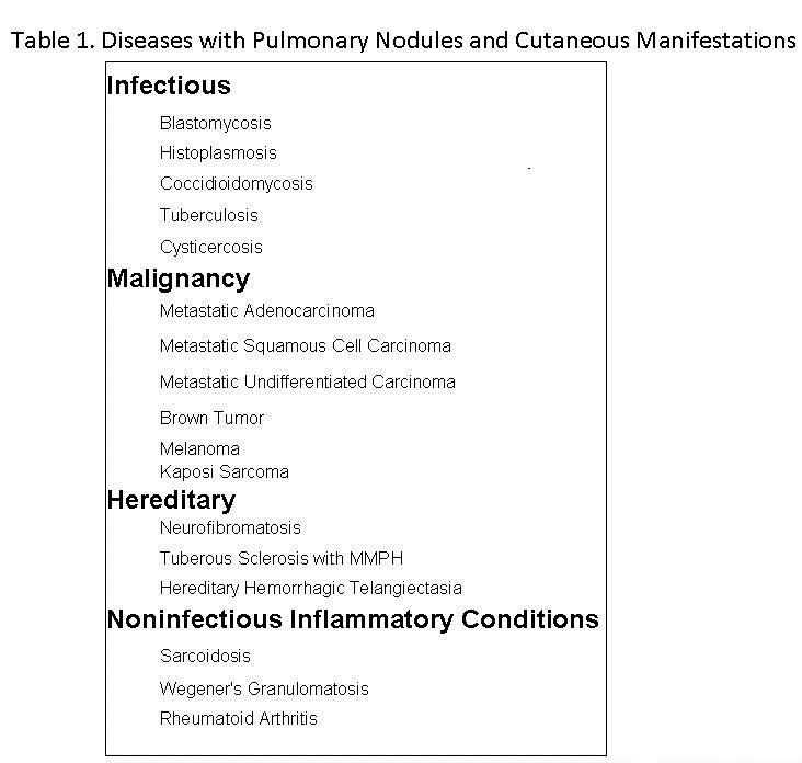
The findings of extrapulmonary nodules in other organ systems and biopsy of nodules can help establish the diagnosis or limit the differential diagnosis of multiple pulmonary nodules when thoracic image findings are nonspecific.

Cysticercosis refers to infection by the larval stage of the pork tapeworm, Taenia solium.  Cysticercosis has emerged as a cause of severe neurologic disease in the United States that primarily affects immigrants from endemic regions.  Within the US cases of cysticercosis are mostly reported in CA, IL, OR, TX, and NY.  Disseminated cysticercosis is an uncommon manifestation of the disease with fewer than 50 cases described worldwide, most occurring in India.  Even more unusual is involvement of the lung parenchyma (1), with less than 10 cases described in literature.  To our knowledge, this is only the second case of disseminated cysticercosis with pulmonary involvement described in North America (2). 

Cysticercosis most commonly affects the central nervous system (neurocysticercosis). However, when disseminated, it frequently involves the skin and subcutaneous tissue.  Cutaneous cysticerci are often a clue to the involvement of internal organs.  In one case series of thirty-three patients with disseminated cysticercosis, sixteen (48%) presented with cutaneous lesions (3).  Detection of the parasite in a biopsy specimen of skin nodules will aid in the diagnosis of disseminated cysticercosis and may prevent further, unnecessary diagnostic tests from being performed.

Sarcoidosis is a common cause of pulmonary nodules with extrapulmonary manifestations.  Recent reports have characterized the development of sarcoidosis in patients receiving pegylated interferon alfa and ribavirin for the treatment of Hepatitis C (4).  In sarcoidosis, there is a predominance of Th1 type immune response, while Th2 lymphocytes are relatively inactivated in granuloma formation (5).  Both IFN-α and ribavirin stimulate the differentiation of Th1-type lymphocytes while inhibiting the activation of Th2-type lymphocytes (6).  Together, this combination therapy works in favor of granuloma formation and the activation/re-activation of sarcoidosis. The temporal association between the initiation of therapy for hepatitis C and the onset of symptoms in the above case raised concern for drug induced sarcoidosis.  However, sarcoidosis is a diagnosis of exclusion and the specific identification of the parasite in the subcutaneous nodule biopsy makes sarcoidosis unlikely. 

The skin is the most common site for disseminated coccidioidomycosis.  Multiple pulmonary nodules with cutaneous lesions in an individual living in Arizona should raise suspicion for coccidioidomycosis. The above patient had a negative coccidioidomycosis work-up and biopsies were not consistent with that of coccidioidomycosis.

Conclusion

Pulmonary cysticercosis is an uncommon manifestation of cysticercosis.  The differential diagnosis of multiple pulmonary nodules is large. However, the diagnosis may be aided by recognizing extrapulmonary lesions that are often associated with lung diseases.  Disseminated cysticercosis with pulmonary involvement should be suspected in any patient presenting with multiple pulmonary nodules who is an immigrant from an endemic region or an individual who has resided in one of the States where cysticercosis is most commonly encountered.

References

  1. Mamere AE, Muglia VF. Disseminated Cysticercosis With Pulmonary Involvement. J Thorac Imaging 2004;19:109-111.
  2. Walts AE, Nivatpumin T, Epstein A. Pulmonary cysticercus. Mod Pathol 1995;8:299-302
  3. Arora PN, Sanchetee PC, Ramakrishnan KR, Venkataram S. Cutaneous, mucocutaneous and neurocutaneous cysticercosis. Indian J Dermatol Venereol Leprol 1990;56:115-8
  4. Ramos-Casals M, Mana J, Nardi N et al. Sarcoidosis in patients with chronic hepatitis C virus infection: analysis of 68 cases. Medicine 2000;84:69-80.
  5. Rodríguez-Lojo, M. Almagro, J. M. Barja, et al., “Subcutaneous Sarcoidosis during Pegylated Interferon Alfa and Ribavirin Treatment for Chronic Hepatitis C,” Dermatology Research and Practice, vol. 2010, Article ID 230417, 2010. doi:10.1155/2010/230417
  6. Tam RC, Pai B, Bard J, et al. Ribavirin polarizes human T cell responses towards a type 1 cytokine profile. J Hepatol 1999;30:376-382.

Address inquires to: Christopher.Strawter@mihs.org

Reference as: Strawter C,  Quiroga P, Zaidi S, Ardiles T. Pulmonary nodules with cutaneous manifestations: A  case report and discussion. Southwest J Pulm Crit Care 2012;4:116-21. (Click here for a PDF version of the manuscript)

Tuesday
Apr032012

March 2012 Pulmonary Case of the Month: There’s Air in There

Alexis Christie, MBBS

Lewis J. Wesselius, MD

History of Present Illness

A 40 year old male was seen with a one week history of dyspnea, dry cough, weakness and abdominal pain. He has a history of acute myelogenous leukemia (AML) diagnosed in December, 2010. He underwent consolidation chemotherapy but had a complication of acute lung injury following chemotherapy thought either to be due to infection or ara-C lung toxicity. Bronchoalveolar lavage was negative and video-assisted thoracotomy revealed only organizing pneumonia.

He underwent stem cell transplantation in May 2011 from a hepatitis C +, allogenic bone marrow transplant and received lamivudine post transplant because of the hepatitis C. Unfortunately, bone marrow biopsy in June 2011 revealed recurrent AML He received two cycles of decitabine.

He had further complications of severe graft versus host disease affecting his eyes, mouth and liver and severe, recurrent C. difficile sepsis. Present medications included: co-trimoxazole (Bactrim), lamivudine, acyclovir, posaconazole, tacrolimus, and prednisone.

Physical exam

Physical exam revealed a thin, moderately short of breath man but was otherwise unremarkable.

Radiology

His chest X-ray (Figure 1) and selected images from his CT scan (Figure 2) are shown below:


Figure 1. Chest x-ray.


Figure 2. Thoracic CT scan. Lung windows.

In addition to the confluent areas of airspace and ground glass opacities throughout both lungs what other finding is present?

  1. Enlarged mediastinal lymph nodes
  2. Pneumothorax
  3. Atelectasis of the left lower lobe
  4. Pneumomediastinum
  5. Large RLL lung mass

Reference as: Christie A, Wesselius LJ. March 2012 pulmonary case of the month: there's air in there. Southwest J Pulm Crit Care 2012;4:88-93. (Click here for a PDF version of the case presentation)

Thursday
Mar222012

Treatment of Coccidioidomycosis-associated Eosinophilic Pneumonia with Corticosteroids

Joshua Malo, MD

Yuval Raz, MD 

Linda Snyder, MD

Kenneth Knox, MD


University of Arizona Medical Center

Department of Medicine

Section of Pulmonary, Allergy, Critical Care, and Sleep Medicine

Tucson, AZ 85724


Abstract

Pulmonary coccidioidomycosis is a common cause of community-acquired pneumonia in endemic areas of the southwestern United States. The clinical spectrum of this disease ranges from an asymptomatic presentation to severe disease with ARDS and hypoxemic respiratory failure. Despite evidence supporting the use of corticosteroids for severe pulmonary disease in other fungal infections, there is currently no established role for this therapy in coccidioidomycosis infections. Peripheral eosinophilia is a common feature of coccidioidomycosis; however, pulmonary eosinophilia is rarely reported. In the setting of pulmonary eosinophilia of other etiologies, corticosteroid therapy has been demonstrated to have a role in reducing the inflammatory response and leading to a more rapid resolution of hypoxemic respiratory failure. We report a case of a patient with primary pulmonary coccidioidomycosis complicated by severe pulmonary eosinophilia that demonstrated rapid improvement after the initiation of corticosteroid therapy.

Case Report

A 71-year-old man presented to the emergency room in Tucson, Arizona with a one-week history of fever, cough, and malaise. The patient’s symptoms began while returning from a trip to northern California. A chest radiograph ordered by the primary care physician demonstrated a right upper lobe consolidation (Figure 1) and azithromycin was prescribed. Fevers persisted along with worsening cough over the next three days, and the patient presented for further evaluation.

Figure 1. Admission radiograph demonstrating right upper lobe airspace disease

Medical history was remarkable for viral cardiomyopathy requiring placement of an ICD after an episode of sudden cardiac death in 2006. An episode of S. bovis bacteremia occurred 4 months prior to the current presentation and was treated with a course of cefazolin. There is no known personal or family history of atopic disease. There is no history of tobacco use or significant occupational exposures. The patient had been living in Arizona during the preceding year and had no other recent travel history, dust, or environmental exposures.

On physical exam, temperature was 38.8°C and pulse oximetry saturation was 90 percent on room air. The patient was in moderate respiratory distress with rales auscultated in the right upper lung zone. Subsequent laboratory examination revealed a PaO2 of 69 mmHg on 4 liters-per-minute of oxygen via nasal cannula. A metabolic panel showed elevated transaminases and his initial leukocyte count was 11.8 x 10³/mL with differential including 5% eosinophils.

The patient was admitted to the medical ward and treated with vancomycin, cefepime, and moxifloxacin for pneumonia caused by a potentially resistant organism. Fluconazole was started on the third hospital day for empiric treatment of primary pulmonary coccidioidomycosis. A CT angiogram of the chest showed bilateral multilobar pneumonia (Figure 2).

Figure 2. CT angiogram of the chest demonstrating multilobar consolidation of the right lung

The patient deteriorated and required intubation for severe hypoxemia two days later. A bronchoalveolar lavage revealed Coccidioides spherules on cytological examination. Liposomal amphotericin B was initiated, which led to the development of oliguric renal failure necessitating hemodialysis. Initial Coccidioides serology was negative, however sputum and BAL cultures demonstrated C. immitis. Despite antifungal therapy his pulmonary status worsened with progressive bilateral pulmonary infiltrates and worsening hypoxemic respiratory failure (Figure 3).

Figure 3. CXR demonstrating progressive bilateral alveolar opacities consistent with ARDS.

In addition, he had a steadily increasing peripheral eosinophilia reaching a maximum of 40 percent with a leukocyte count of 14.8 x 10³/mL despite the absence of any signs of disseminated coccidioidomycosis. A repeat BAL again showed Coccidioides spherules and eosinophils of 40 and 56 percent from the right middle lobe and lingula, respectively. Methylprednisolone 40mg IV three times daily was started with a decline in blood eosinophils to one percent within 24 hours. Chest radiographs and A-a gradient rapidly improved over the next 3 days leading to successful extubation. The patient was transitioned to oral fluconazole and prednisone and discharged from the hospital in good condition two weeks later.

At the follow-up six weeks after initial presentation, he remains on fluconazole and prednisone 15mg daily with no signs of disseminated coccidioidomycosis and is continuing a gradual reduction of prednisone dosage.

Discussion

Coccidioidomycosis is caused by either of 2 species of the dimorphic fungus Coccidioides. Endemic regions are present in North and South America, with the majority of cases within the United States arising in Arizona and California. Although peripheral eosinophilia is a commonly reported finding (1), pulmonary eosinophilia has rarely been described.

Acute eosinophilic pneumonias may be idiopathic or a secondary inflammatory response to various infections or environmental exposures. In regions where endemic fungal infections are common, differentiating between eosinophilic pneumonias of idiopathic versus infectious etiology is vital in order to avoid inappropriate therapy and its adverse consequences. A review of the literature concerning pulmonary coccidioidomycosis and concurrent pulmonary eosinophilia demonstrates only 9 prior case reports. Corticosteroid therapy was used for treatment of the pulmonary eosinophilia in only 3 of these cases, 2 of which resulted in death from disseminated coccidioidal infection (1-3). One case ended in spontaneous resolution of disease without antifungals or corticosteroids leading the authors to suggest a conservative approach with corticosteroids due to the risk for dissemination (4).

In our case, there was progressive clinical deterioration despite ten days of treatment with appropriate antifungal regimen, leading to our decision to treat with corticosteroids. The immediate decrease in peripheral eosinophilia in conjunction with the rapid clinical improvement leads us to the conclusion that corticosteroids were beneficial in the resolution of his acute respiratory failure. The clinical response observed is similar to that expected in idiopathic acute eosinophilic pneumonia which supports the notion that the eosinophilic response, as opposed to the primary infection, was primarily responsible for our patient’s severe hypoxemia.

There remains a risk for disseminated disease. In the cases cited in which patients died of dissemination, antifungal therapy preceding corticosteroid therapy was not described. Due to the risk of underlying pulmonary coccidioidomycosis in endemic regions, corticosteroid therapy for eosinophilic pneumonia should only be considered in the setting of severe hypoxemic respiratory failure and once adequate antifungal therapy has been initiated.

According to recent guidelines there is no role for corticosteroid therapy in the treatment of coccidioidomycosis due to a lack of convincing data for efficacy and safety (5). There is precedent for treating severe pulmonary disease caused by other fungal infections, such as histoplasmosis and blastomycosis, with corticosteroids. We suggest that there is a role for the use of corticosteroid therapy in the setting of progressive respiratory failure due to coccidioidomycosis with associated pulmonary eosinophilia that has failed conventional antifungal therapy.

References

  1. Echols RM, Palmer DL, Long GW. Tissue eosinophilia in human coccidioidomycosis. Rev Infect Dis 1982;4:656–664.
  2. Lombard CM, Tazelaar HD, Krasne DL. Pulmonary eosinophilia in coccidioidal infections. Chest 1987;5:734–736
  3. Swartz J, Stoller JK. Acute Eosinophilic Pneumonia Complicating Coccidioides immitis Pneumonia: A Case Report and Literature Review. Respiration 2009;77:102–106
  4. Whitlock WL, Dietrich RA, Tenholder MF. Acute eosinophilic pneumonia (letter). N Engl J Med 1990;322:635
  5. Limper AH, Knox KS, Sarosi GA, et al. Treatment of fungal infections in adult pulmonary and critical care patients. Am J Respir Crit Care Med 2011;183:96–128

The authors report no conflicts of interest

Address correspondence to:     Joshua Malo, MD

                                             University of Arizona Medical Center

                                             Department of Medicine    

                                             Section of Pulmonary, Allergy, Critical

                                             Care and Sleep Medicine

                                             Tucson, AZ 85724

                                             E-mail: jmalo@deptofmed.arizona.edu


Reference as: Malo J, Raz Y, Snyder L, Knox K. Treatment of coccidioidomycosis-associated eosinophilic pneumonia with corticosteroids. Southwest J Pulm Crit Care 2012;4:61-66. (Click here for a PDF version of the manuscript) 

Wednesday
Mar072012

Sympathetic Empyema Arising from Streptococcus anginosus Splenic Abscess

Emad Wissa, MD

Robert A. Raschke, M.D.

Manoj Mathew, MD, FCCP, MCCM 

Good Samaritan Regional Medical Center

1111 E. McDowell Road

Phoenix, AZ 85006


Abstract

We report a 52 year old male with a history splenic infarction, abdominal pain and shortness of breath. CT scanning revealed a splenic abcess and empyema. Cultures from both sites grew Streptococcus anginosus. These resolved with drainage and antibiotics. Physicians should consider Streptococcus species when confronted with a patient with splenic infarction.

Introduction

Sympathetic Empyema Thoracis or sympathetic empyema is a rare entity. The mechanism by which this occurs is via penetration by a subdiaphragmatic abscess into the pleural space. Most reported cases have occurred in the right hemithorax secondary to the presence of a liver abscess or ascites. Splenic abscess are rare and are often seen in the setting of embolic endocarditis, infections from Salmonella or other bacterial invade the infarcted spleen (1,2). To our knowledge, this is the first reported case of sympathetic empyema likely secondary to a Streptococcus splenic abscess.

Case Report

A 52 year old male presented with a 2 day history of shortness of breath and 2 a month history of left upper quadrant pain. His medical history included hepatitis C, drug abuse and splenic infarction. On examination his breathing was labored. His chest X-ray was remarkable for a left lower lobe opacification (Figure 1).

Figure 1. Chest x-ray showing left lower lobe opacification.

He was endotracheally intubated and started on broad spectrum antibiotics. A computed tomography (CT) of the chest and abdomen demonstrated a moderate left side pleural effusion, compressive atelectasis, a small amount of ascites and a splenic abscess (Figure 2).

Figure 2. Coronal CT of chest and upper abdomen.


A chest tube was placed into the left hemithorax, and a pigtail catheter was placed into the splenic abscess. Cultures from both sites yielded Streptococcus anginosus. The empyema was treated with antibiotics, and chest tube fibrinolytic therapy with tissue plasminogen activator (TPA) and dornase alpha. Blood cultures were sterile and echocardiogram revealed no vegetations. The patient was extubated on hospital day 4. The left pleural empyema responded well to fibrinolytic therapy with chest tube removal on day 10. The splenic abscess required long term drainage with pigtail remaining in place for 3 weeks before the abscess resolved. The patient was discharged home on hospital day 24 on amoxicillin.

Discussion

Sympathetic empyema is an infection in the pleural space caused by translocation of a bacterial infection from the liver, ascitic fluid or from a splenic abscess. This patient had a known history of splenic infarction predisposing him to bacterial invasion and splenic abscess formation. The absence of cough, sputum production, and lobar consolidation argues against an empyema arising from and underlying pneumonia, although we can not exclude that possibility. Our review of the medical literature demonstrates no known cases of sympathetic empyema from Streptococcus anginosus. Physicians should also consider this organism in a patient with a splenic abscess.

References

  1. Buscaglia A. Empyema due to splenic abscess with Salmonella newport. JAMA1978;240:1990.
  2. Tornos M.P, Mayor G, Nadal A, Soler A. Empyema and splenic abscess in infective endocarditis. Int J Cardio 1984;6:746-8.

Reference as: Wissa E, Raschke RA, Mathew M. Sympathetic empyema arising from streptococcus anginosus splenic abscess. Southwest J Pulm Crit Care 2012;4:48-50. (Click here for a PDF version of the case presentation)

Friday
Feb242012

February 2012 Pulmonary Case of the Month

Lewis J. Wesselius, MD

Associate Editor Pulmonary

History of Present Illness

A 49 year old female was seen for fever and shortness of breath in December 2011. She has a history of right infiltrating ductal breast cancer diagnosed in 2001 at age 38. Treatment included mastectomy with negative lymph nodes, followed by 4 courses of doxarubicin and cytoxan. She did well until March 2010 when erythema was noted over her right chest.  Biopsy showed adenocarcinoma consistent with  breast carcinoma. The biopsy was “triple negative”, i.e., negative for estrogen receptors, progesterone receptors and Her2. PET scan demonstrated multiple positive lymph nodes in the mediastinum, supraclavicular area and bone metastases. She received radiation to her chest wall and chemotherapy most recently gemcitabine, carboplatin and iniparib. In October 2011 brain metastases were noted and she was started on stereotactic brain radiation and dexamethasone.

Past Medical, Family and Social Histories

  • She had a prothrombin mutation noted and was begun prophylactically on warfarin in 2010.
  • There was a family history of breast carcinoma.
  • She was a non-smoker with no unusual exposures.
  • Current medications include omeprazole, metoprolol, and warfarin.

Physical Examination

She was receiving oxygen at 3 L/min by nasal cannula. Temperature was to 37.9°C. She had bilateral crackles on chest auscultation, most prominent at bases. Physical Examination was otherwise noncontributory.

Initial Laboratory Evaluation

  • Hemoglobin/Hematocrit 11.8 g/dL/33.9%
  • White blood count 5.1 X10⁹/L
  • Platelets 64 X 109/L
  • INR 1.58 

Chest CT scan

Chest CT scan is in figure 1.

Figure 1. Selected images from the admission CT scan. The CT scan was interpreted as showing diffuse groundglass opacities and scattered centrilobular nodules. 

Which of the following diagnosis are consistent with the patient’s presentation and CT scan?

  1. Pulmonary edema
  2. Bacterial pneumonia
  3. Fungal pneumonia
  4. Drug reaction
  5. All of the above

Reference as: Wesselius LJ. February 2012 pulmonary case of the month. Southwest J Pulm Crit Care 2012;4:42-7.

(Click here for a PDF version of the case presentation)

(Click here for a Powerpoint slide presentation of the case)