Search Journal-type in search term and press enter
Southwest Pulmonary and Critical Care Fellowships
Social Media

Pulmonary

Last 50 Pulmonary Postings

(Click on title to be directed to posting, most recent listed first)

June 2025 Pulmonary/Critical Care Case of the Month: Hemoptysis
   from a Very Unusual Cause
March 2025 Pulmonary Case of the Month: Interstitial Lung Disease of
   Uncertain Cause
December 2024 Pulmonary Case of the Month: Two Birds in the Bush Is
   Better than One in the Hand
Glucagon‐like Peptide-1 Agonists and Smoking Cessation: A Brief Review
September 2024 Pulmonary Case of the Month: An Ounce of Prevention
   Caused a Pound of Disease
Yield and Complications of Endobronchial Ultrasound Using the Expect
   Endobronchial Ultrasound Needle
June 2024 Pulmonary Case of the Month: A Pneumo-Colic Association
March 2024 Pulmonary Case of the Month: A Nodule of a Different Color
December 2023 Pulmonary Case of the Month: A Budding Pneumonia
September 2023 Pulmonary Case of the Month: A Bone to Pick
A Case of Progressive Bleomycin Lung Toxicity Refractory to Steroid Therapy
June 2023 Pulmonary Case of the Month: An Invisible Disease
February 2023 Pulmonary Case of the Month: SCID-ing to a Diagnosis
December 2022 Pulmonary Case of the Month: New Therapy for Mediastinal
   Disease
Kaposi Sarcoma With Bilateral Chylothorax Responsive to Octreotide
September 2022 Pulmonary Case of the Month: A Sanguinary Case
Electrotonic-Cigarette or Vaping Product Use Associated Lung Injury:
   Diagnosis of Exclusion
June 2022 Pulmonary Case of the Month: A Hard Nut to Crack
March 2022 Pulmonary Case of the Month: A Sore Back Leading to 
   Sore Lungs
Diagnostic Challenges of Acute Eosinophilic Pneumonia Post Naltrexone
   Injection Presenting During The COVID-19 Pandemic
Symptomatic Improvement in Cicatricial Pemphigoid of the Trachea
   Achieved with Laser Ablation Bronchoscopy
Payer Coverage of Valley Fever Diagnostic Tests
A Summary of Outpatient Recommendations for COVID-19 Patients
   and Providers December 9, 2021
December 2021 Pulmonary Case of the Month: Interstitial Lung
   Disease with Red Knuckles
Alveolopleural Fistula In COVID-19 Treated with Bronchoscopic 
   Occlusion with a Swan-Ganz Catheter
Repeat Episodes of Massive Hemoptysis Due to an Anomalous Origin 
   of the Right Bronchial Artery in a Patient with a History
   of Coccidioidomycosis
September 2021 Pulmonary Case of the Month: A 45-Year-Old Woman with
   Multiple Lung Cysts
A Case Series of Electronic or Vaping Induced Lung Injury
June 2021 Pulmonary Case of the Month: More Than a Frog in the Throat
March 2021 Pulmonary Case of the Month: Transfer for ECMO Evaluation
Association between Spirometric Parameters and Depressive Symptoms 
   in New Mexico Uranium Workers
A Population-Based Feasibility Study of Occupation and Thoracic 
   Malignancies in New Mexico
Adjunctive Effects of Oral Steroids Along with Anti-Tuberculosis Drugs
   in the Management of Cervical Lymph Node Tuberculosis
Respiratory Papillomatosis with Small Cell Carcinoma: Case Report and
   Brief Review
December 2020 Pulmonary Case of the Month: Resurrection or 
   Medical Last Rites?
Results of the SWJPCC Telemedicine Questionnaire
September 2020 Pulmonary Case of the Month: An Apeeling Example
June 2020 Pulmonary Case of the Month: Twist and Shout
Case Report: The Importance of Screening for EVALI
March 2020 Pulmonary Case of the Month: Where You Look Is 
   Important
Brief Review of Coronavirus for Healthcare Professionals February 10, 2020
December 2019 Pulmonary Case of the Month: A 56-Year-Old
   Woman with Pneumonia
Severe Respiratory Disease Associated with Vaping: A Case Report
September 2019 Pulmonary Case of the Month: An HIV Patient with
   a Fever
Adherence to Prescribed Medication and Its Association with Quality of Life
Among COPD Patients Treated at a Tertiary Care Hospital in Puducherry
 – A Cross Sectional Study
June 2019 Pulmonary Case of the Month: Try, Try Again
Update and Arizona Thoracic Society Position Statement on Stem Cell 
   Therapy for Lung Disease
March 2019 Pulmonary Case of the Month: A 59-Year-Old Woman
   with Fatigue
Co-Infection with Nocardia and Mycobacterium Avium Complex (MAC) 
   in a Patient with Acquired Immunodeficiency Syndrome 
Progressive Massive Fibrosis in Workers Outside the Coal Industry: A Case 
   Series from New Mexico
December 2018 Pulmonary Case of the Month: A Young Man with
   Multiple Lung Masses
Antibiotics as Anti-inflammatories in Pulmonary Diseases
September 2018 Pulmonary Case of the Month: Lung Cysts
Infected Chylothorax: A Case Report and Review
August 2018 Pulmonary Case of the Month
July 2018 Pulmonary Case of the Month
Phrenic Nerve Injury Post Catheter Ablation for Atrial Fibrillation
Evaluating a Scoring System for Predicting Thirty-Day Hospital
   Readmissions for Chronic Obstructive Pulmonary Disease Exacerbation


For complete pulmonary listings click here.

The Southwest Journal of Pulmonary, Critical Care & Sleep publishes articles broadly related to pulmonary medicine including thoracic surgery, transplantation, airways disease, pediatric pulmonology, anesthesiolgy, pharmacology, nursing  and more. Manuscripts may be either basic or clinical original investigations or review articles. Potential authors of review articles are encouraged to contact the editors before submission, however, unsolicited review articles will be considered.

-------------------------------------------------------------------------------------

Monday
Mar032014

Elevated Tumor Markers In Coccidioidomycosis of the Female Genital Tract

Ying Wu, MD1

Stacy Arnold2

Tim Kuberski, MD3


1Department of Internal Medicine and the 3Section of Infectious Disease

Maricopa Medical Center

Phoenix, Arizona

2University of Arizona College of Medicine Phoenix

Phoenix, Arizona


Abstract

The female genital tract is rarely involved by coccidioidomycosis. We describe a woman with disseminated coccidioidomycosis involving the female pelvic organs associated with elevated tumor markers CA 125 and CA 19-9. She had no fevers and the initial clinical suspicion was a malignancy because of the elevated tumor markers. At exploratory laparotomy a total abdominal hysterectomy and bilateral salpingo-oophorectomy were performed because of the suspicion of a malignancy. Subsequent pathology demonstrated coccidioidomycosis involving the female genital tract and no malignancy. The abnormal CA 125 and CA 19-9 returned to normal after surgical resection and treatment of the coccidioidomycosis.

Introduction

Dissemination of coccidioidomycosis to the female genital tract is an unusual complication of that fungal infection (1). This report describes a woman with disseminated coccidioidomycosis which mimicked a malignancy of the female genital tract. The initial clinical suspicion was a malignancy because of the elevation of carbohydrate antigen tumor markers CA125 and CA19-9.

Case Report

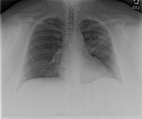
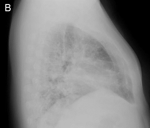
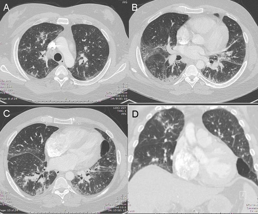
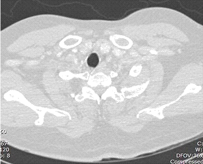
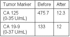
A 48-year-old Hispanic woman with a history of adult onset diabetes and menorrhagia presented with left lower quadrant pain.  She denied fever, chills or sweats.  She gave no history of “Valley Fever” or pneumonia.  Physical examination was significant for left lower quadrant abdominal tenderness without rebound or guarding.  CT of the abdomen and pelvis showed free fluid within the pelvis, masses within the uterus, left hydronephrosis and bilateral cystic masses of the adnexa. She was anemic and thrombocytopenic with a hemoglobin of 7.8 g/dL and platelet count of 51,000/mm3.  She received blood and platelet transfusions and was given oral prednisone 80 mg once daily for five days which was associated with improvement of her platelet count to normal. Certain tumor markers were found to be elevated a CA 125 of 475.7 (normal range: 0-35 U/mL) and CA 19-9 of 133 (normal range: 0–37 U/mL). A carcinoembryonic antigen (CEA) was not elevated 1.9 (normal range 0-2.5 ng/ml). Her diabetes was controlled. The left hydronephrosis was treated with a ureteral stent and nephrostomy.  An endometrial biopsy was consistent with benign endocervical and endometrial tissue.  Percutaneous drainage of the left ovarian cyst fluid showed no malignant cells and no abscess.  A plain chest x-ray was interpreted as normal, however a CT scan of the chest showed a left lower lobe lesion which was biopsied and found to be consistent with an old Coccidioides granuloma.  Because of the concern for a pelvic malignancy, an exploratory laparotomy was done and a total hysterectomy with bilateral salpingo-oophorectomy was performed.  The pathology of the surgical specimen showed Coccidioides with necrotizing granulomas and spherules in the round ligament, uterus, endometrium, bilateral ovaries and fallopian tubes (Figure 1).

Figure 1.  Hematoxylin and eosin stain of round ligament showing necrotizing granulomas associated with Coccidioides spherules (arrows). Empty Coccidioides  spherule in inset.

Following the operation, she was started on oral fluconazole 400mg daily and followed as an outpatient. Her complement fixation titer for Coccidioides was positive at 1:64 on discharge, increased to 1:256 one month later and then declined to 1:8 five months later. At five months follow-up she was asymptomatic and back to her premorbid state. She was to remain on fluconazole indefinitely.  Postoperatively the CA 125 and CA 19-9 returned to normal at 12.3 and 12.0 respectively after fifty days (Table 1). 

Table 1. Tumor Marker Values Before and Fifty Days After Surgical Resection.


Discussion 

Dissemination of Coccidioides to the female pelvic organs is unusual. In a series of 142 necropsies of patients who died of coccidioidomycosis, none of the thirty-three females had involvement of the genital tract (2).  Presumptively the fungus spreads endogenously from the lung as in our patient. It is notable that the lung lesion in this patient was initially thought to be a malignancy. However, when “old Coccidioides granuloma” was reported, it was not associated with her genital tract problem by the treating physicians. We were able to find seventeen previously reported cases of coccidioidomycosis involving the female genital tract since the first report in 1929 (3-7). In those cases of Coccidioides involving the genital tract, there was no combination of investigations or clinical features that were sensitive enough to diagnose the female genital tract infection preoperatively. In fourteen of the eighteen patients (including our patient) with Coccidioides serologic test results, thirteen had a positive titer of 1:16 or greater (3-6).

In all eighteen cases the diagnosis of Coccidioides involving the genital tract was made only post-operatively after the microscopic examination of surgical specimens. Treatment of female genital tract coccidioidomycosis generally requires surgical resection of the involved tissues and antifungal agents (3). Cure of pelvic coccidioidomycosis by surgical resection alone, without antifungal therapy, has been reported in at least 5 cases (3,7). However, deaths attributable to disseminated coccidioidomycosis with involvement of the genital tract despite treatment and surgery have been reported (8). Quantitative serologic testing can be useful in making the diagnosis and following the response to treatment. The complement fixation titers to Coccidioides would be expected to decrease with treatment and clinical improvement. Our patient’s complement fixation titer went from a high of 1:256 to 1:8 after surgical resection and five months of treatment. In general, patients with disseminated coccidioidomycosis will respond to fluconazole, but the duration of therapy can be protracted for years.

Early in the course of our patient’s workup, she was found to have elevated CA 125 and CA 19-9 levels, raising the suspicion of a malignancy. In general, tumor markers should not be used as a screening test for malignancy and are most useful for following the response to treatment. In this patient the response was to antifungal therapy rather than chemotherapy. We believe the tumor markers were elevated due to the inflammatory response to the Coccidioides infection (4,9). CA 125 is most commonly monitored in the management of ovarian carcinoma.  However, it is only 75-80% sensitive in patients with ovarian cancer and can also be elevated in a variety of benign conditions such as infection (10).  In addition to the ovarian cancer cells, benign endometrial and peritoneal mesothelial cells can also secrete CA 125 (11). It appears that any process causing serosal inflammation may elevate CA 125. Thus non-tumorous conditions such as pelvic tuberculosis can result in sufficient inflammation to cause an increased CA 125 level (12). CA 19-9 is a serum marker associated with pancreatic cancer, but it also can be elevated in non-cancerous conditions such as pancreatitis, endometriosis and in diabetics with poor glycemic control (10,13).

Both CA 125 and CA 19-9 can be elevated in noncancerous conditions such as infection and presumed to be the reason for the elevation in our patient. Her surgical histopathology showed concomitant endometriosis of the round ligament, uterine serosa and both ovaries (Figure 2).

Figure 2. Hematoxylin and eosin stain of ovary showing changes consistent with endometriosis (arrow).

This was in addition to the necrotizing granulomas containing Coccidioides spherules (Figure 1). Increased levels of CA 125 have been reported previously in three patients with Coccidioides involving pelvic organs (4,5,7), but there have been no reports of elevated serum CA 19-9 levels.  Endometriosis alone can cause both CA 125 and CA 19-9 to be increased (14,15), however the mechanism by which these markers become elevated is not completely understood. It has been postulated that chronic inflammation causes an epithelial reaction that results in the leakage of the tumor markers into the circulation (14,15).  Endometriosis due to a variety of infectious etiologies can cause serum CA 125 levels to be elevated and they can precipitously decrease following surgical intervention, as in our patient (4,5). CA 19-9 levels have also been found to return to normal following surgical intervention for endometriosis (16). We presume that the elevated tumor markers in our patient were the result of similar inflammatory mechanisms observed for both CA 125 and CA 19-9. We suspect the reason that CA 19-9 levels have not been reported previously is that this test would be ordered less frequently in patients suspected of having a pelvic malignancy. Our patient had a normal CEA level consistent with a lack of involvement of the gastrointestinal tract by coccidioidomycosis.

Another potential source of Coccidioides infecting the female genital tract is sexual transmission. We could find no references to document sexual transmission of Coccidioides. However, transmission from a man with prostate involvement to a woman who developed an infection of her genital tract could occur, but our patient’s husband had no prostate disease, no history of Valley Fever and his Coccidioides serology was negative. Coccidioides has been documented to mimic a variety of malignancies such as breast cancer, bronchogenic carcinoma, osteosarcoma and lymphoma (6). It is notable that the first reported case of coccidioidomycosis in 1892 was initially misdiagnosed as mycosis fungoides (6). Coccidioidomycosis is known to have protean manifestations and in this patient it mimicked a female genital tract malignancy. This case is unique because a Coccidioides infection was documented and associated with the elevation of tumor markers CA 19-9 and CA 125, which returned to normal following treatment and surgical resection of the involved tissues. Involvement of the female genital tract by coccidioidomycosis may be uncommon, but in endemic areas it should be a consideration in the differential diagnosis in women with a suspected pelvic malignancy and elevated levels of CA 125 and CA 19-9.

References

  1. Saw EC, Smale LE, Einstein H, Huntington RW,Jr. Female genital coccidioidomycosis. Obstet Gynecol. 1975;45:199-202. [PubMed] 
  2. Huntington RW Jr., Waldmann WJ, Sargent JA, O'Connell H, Wybel R, Croll D. pathological and clinical observations in 142 cases of fatal coccidioidomycosis on necropsy. In Ajello L, ed. Proceedings of the Second Coccidioidomycosis Symposium. Tuscon, The University of Arizona Press.1967:143-167.
  3. Chowfin A, Tight R. Female genital coccidioidomycosis (FGC), Addison's disease and sigmoid loop abscess due to Coccidioides immites; case report and review of literature on FGC. Mycopathologia. 1999;145:121-126. [CrossRef] [PubMed]
  4. Ellis MW, Dooley DP, Sundborg MJ, Joiner LL, Kost ER. Coccidioidomycosis mimicking ovarian cancer. Obstet Gynecol. 2004;104:1177-1179. [CrossRef] [PubMed] 
  5. Micha JP, Goldstein BH, Robinson PA, Rettenmaier MA, Brown JV. Abdominal/pelvic Coccidioidomycosis. Gynecol Oncol. 2005;96:256-258. [CrossRef] [PubMed]
  6. Crum-Cianflone NF, Truett AA, Teneza-Mora N, et al. Unusual presentations of coccidioidomycosis: a case series and review of the literature. Medicine (Baltimore). 2006;85:263-277. [CrossRef] [PubMed] 
  7. Smith G, Hoover S, Sobonya R, Klotz SA. Abdominal and pelvic coccidioidomycosis. Am J Med Sci. 2011;341:308-311. [CrossRef] [PubMed]
  8. Bylund DJ, Nanfro JJ, Marsh WL,Jr. Coccidioidomycosis of the female genital tract. Arch Pathol Lab Med. 1986;110:232-235. [PubMed] 
  9. Bast RC,Jr, Xu FJ, Yu YH, Barnhill S, Zhang Z, Mills GB. CA 125: the past and the future. Int J Biol Markers. 1998;13:179-187. [PubMed] 
  10. Cancer antigen 125. In: Ferri FF, ed. Ferri's Clinical Advisor 2013 : 5 Books in 1. 1st ed. St. Louis, Mo: Elsevier Mosby; 2013:1452-1453.
  11. Sevinc A, Camci C, Turk HM, Buyukberber S. How to interpret serum CA 125 levels in patients with serosal involvement? A clinical dilemma. Oncology. 2003;65:1-6. [CrossRef] [PubMed] 
  12. Sheth SS. Elevated CA 125 in advanced abdominal or pelvic tuberculosis. Int J Gynaecol Obstet. 1996;52:167-171. [CrossRef] [PubMed]
  13. Yu H, Li R, Zhang L, Chen H, Bao Y, Jia W. Serum CA19-9 level associated with metabolic control and pancreatic beta cell function in diabetic patients. Exp Diabetes Res. 2012;2012:745189. [CrossRef] [PubMed] 
  14. Kurata H, Sasaki M, Kase H, Yamamoto Y, Aoki Y, Tanaka K. Elevated serum CA125 and CA19-9 due to the spontaneous rupture of ovarian endometrioma. Eur J Obstet Gynecol Reprod Biol. 2002;105:75-76. [CrossRef]  [PubMed]
  15. Park BJ, Kim TE, Kim YW. Massive peritoneal fluid and markedly elevated serum CA125 and CA19-9 levels associated with an ovarian endometrioma. J Obstet Gynaecol Res. 2009;35:935-939. [CrossRef] [PubMed] 
  16. Takemori M, Sugimura K. Ovarian chocolate cyst with markedly elevated serum CA19-9 level: a case report. Eur J Obstet Gynecol Reprod Biol. 1991;42:241-244. [CrossRef] [PubMed] 

Reference as: Wu Y, Arnold S, Kuberski T. Elevated tumor markers in coccidiomyocosis of the female genital tract. Southwest J Pulm Crit Care. 2014;8(3):170-5. doi: http://dx.doi.org/10.13175/swjpcc179-13 PDF

Saturday
Mar012014

March 2014 Pulmonary Case of the Month: The Cure May Be Worse Than the Disease

Sudheer Penupolu, MD 

Philip J. Lyng, MD

Lewis J. Wesselius, MD 


Department of Pulmonary Medicine

Mayo Clinic Arizona

Scottsdale, AZ


History of Present Illness

A 51 year old woman was seen with a chief complaint of gradually increasing shortness of breath. She was at baseline five months prior to presentation but noticed dyspnea on minimal exertion initially at a higher altitude, gradually progressing to dyspnea at rest. She was tried on 2 courses of antibiotics with no significant improvement. In addition to the dyspnea, she has some non productive cough but no fevers.

PMH, SH, FH

She had a renal transplant in 1997 for IgA disease and has a history of type II diabetes and hypertension.

She is a life long nonsmoker and has only occasional alcohol use. She is employed as a utility designer and has no exposure to any dusts, fumes or exotic animals.

Family history is noncontributory.

Medications

  • Atenolol
  • Lasix
  • Prednisone 2 mg q daily
  • Rosuvastatin
  • Sirolimus 2 mg po q daily

There have been no changes in the doses in the past few years.

Physical Examination

Physical examination reveals no abnormalities and her lung auscultation is clear.

Laboratory

Her complete blood count (CBC), urinanalysis, liver function tests, and calcium were all within normal limits.

Radiology

An x-ray of the chest is shown in Figure 1. 

Figure 1. Initial PA chest radiograph.

Which of the below is the best interpretation of her chest x-ray?

  1. Cardiomegaly
  2. Left upper lobe consolidation
  3. Normal
  4. Right upper lobe consolidation
  5. All of the above

Reference as: Penupolu S, Lyng PJ, Wesselius LJ. March 2014 pulmonary case of the month: the cure may be worse than the disease. Southwest J Pulm Crit Care. 2014;8(3):142-51. http://dx.doi.org/10.13175/swjpcc005-14 PDF


Saturday
Feb012014

February 2014 Pulmonary Case of the Month: Faster Is Not Always Better

Lewis Wesselius MD


Department of Pulmonary Medicine

Mayo Clinic Arizona

Scottsdale, AZ


History of Present Illness

A 56 year old woman with a history of rheumatoid arthritis (RA) for 26 years was seen as an outpatient. She has a recent history of increased cough, sputum and dyspnea.

PMH, FH, SH

She was originally from India but had lived in Singapore from 2011 to June 2013 before moving to Phoenix. In 2009, she was diagnosed with Mycobacterium avium-intracellulare (MAI) on bronchoscopy and started on azithromycin, ethambutol, and rifabutin. She was unable to tolerate rifabutin but was continued on ethambutol and azithromycin. She had been on etanercept for her RA which was held after the diagnosis of MAI. She had negative sputum cultures for MAI in September 2012 and her ethambutol and azithromycin were stopped. In May 2013 she had increased symptoms and bronchoscopy demonstrated Pseudomonas and nontuberculous mycobacterium (NTM). She was treated with cefipime/ciprofloxacin for 6 weeks prior to moving to Phoenix.

She does not smoke or drink. Her FH is noncontributory.

Medications

  • Celecoxib 200 mg bid
  • Gabapentin 600 bid
  • Methotrexate 15 mg weekly
  • Prednisone 5 mg daily
  • Tramadol 50 mg every 4 hours prn

Physical Examination

Afebrile. SpO2 96% on room air.

Chest: scattered crackles in both lungs, no wheezes.

There were joint deformities typical of chronic RA, but otherwise the remainder of the physical exam was unremarkable.

Radiology

She brings a CT scan from 2009 (Figure 1). 

Figure 1. Panels A-D: Representative static axial lung images from a thoracic CT scan performed in 2009. Lower panel: movie of selected lung images from the thoracic CT scan performed in 2009.

What should be done next? (Click on correct answer to move to next panel)

  1. Repeat the CT scan
  2. Restart ethambutol and azithromycin
  3. Sputum culture
  4. 1 + 3
  5. All of the above

Reference as: Wesselius LJ. February 2014 pulmonary case of the month: faster is not always better. Southwest J Pulm Crit Care. 2014:8(2):74-8. doi: http://dx.doi.org/10.13175/swjpcc168-13 PDF

Wednesday
Jan012014

January 2014 Pulmonary Case of the Month: Too Much, Too Late

Chidinma Chima-Okereke MD  

Department of Pulmonary Medicine

Cedars Sinai Medical Center

Los Angeles, CA


Chief Complaint: Difficulty breathing

History of Present Illness

A 49-year-old gentleman with history of hepatitis C cirrhosis complicated by ascites presented to the emergency room of Olive View Medical Center in San Fernando Valley, California complaining of worsening shortness of breath. The patient reports that he occasionally has shortness of breath, usually about 2-3 times a year. However for the past 2 months, he has had worsening dyspnea on exertion and cannot walk further than 5 minutes. He also reports orthopnea and paroxysmal nocturnal dyspnea. He has been having a dry cough for the past 3-4 weeks.

He has a history of chronic ascites that has required multiple taps. He has been taking his prescribed diuretics however instead of taking these medications daily he takes them about every other day due to financial constraints.

However, his abdominal distention and his lower extremity swelling are stable. He reports some nausea with decreased appetite. He also has a new symptom of left-sided chest pain that radiates down his left arm and shoulder that lasts about 20 minutes and has no associated symptoms. .

He denies any fevers or chills or weight change. He has no sick contacts.

Past Medical and Surgical History

  • Hepatitis C cirrhosis
  • Chronic lower extremity edema
  • Ascites, status post multiple large volume paracentesis
  • History of chronic abdominal pain treated with morphine
  • Status post chest tube when he was a 17-year-old due to a gunshot wound

Social History

  • History of incarceration, released about 8 months ago
  • 6-pack of beer a day – quit 12 years ago.
  • Former smoker, quit 10 years ago, 7 pack-years
  • IV heroin use 15 years ago
  • No cocaine, amphetamines or any inhaled substances
  • No recent travel, occupational, pet or bird exposures
  • Lives with his fiancé in Lancaster, California

Family History

  • Father died of an MI at age 56.
  • Mother - SLE, DM, Stroke
  • Sister - Colon cancer
  • Brother - Hepatitis C cirrhosis

Medications

  • Controlled-release morphine sulfate 15 mg p.o. every morning and 30 mg p.o. every evening.
  • Furosemide 40 mg p.o. daily.
  • Spironolactone 50 mg p.o. daily.
  • Lactulose 15 mL p.o. b.i.d. p.r.n.

Review of Systems

Positive for pleuritic chest pain, night sweats, chills, dry cough - unproductive of sputum, lightening and darkening of urine, lower extremity edema, palpitations, decreased appetite, dry mouth, joint stiffness in the morning.    

Physical Examination

  • Vital signs: T 97.4 BP 115/67, HR 89, RR 20, SpO2 93%/RA
  • Lung exam was significant for bilateral crackles midway up the back.
  • Abdominal exam was non-tender and not suggestive of ascites
  • Lower extremities: 1+ bilateral pitting edema up to the knees.
  • Multiple skin tattoos and erythema in his lower extremities  
  • Muscle strength was 3/5 in the lower extremities, 4/5 in upper extremities bilaterally.
  • Otherwise the physical exam was unremarkable.

Laboratory

  • Basic Metabolic Panel was within normal limits.
  • Complete blood count (CBC): White count 6.3 X 10³/mm3 with 8.3% eosinophils, hemoglobin 12.3 g/dL, platelets 130,000/µL.
  • Liver function tests (LFTs): AST 78 IU/L, ALT 42 IU/L, alkaline phosphatase 115, total bilirubin 1.3 mg/dL, INR 1.3, albumin 2.7 g/dL.
  • Brain naturetic peptide (BNP) 38 ng/L, troponin is 0.008 ng/ml.

Radiography

A chest x-ray was obtained (Figure 1).

Figure 1. Admission AP (Panel A) and lateral (Panel B) chest x-ray.

The chest x-ray was interpreted as poor inspiration with elevation of the right diaphragm. The heart is at least upper limits of normal in size. Pulmonary vessels are congested. The azygos vein is mildly dilated. No significant pleural effusion is detected in these two views.

A CT angiogram was obtained to rule out pulmonary embolism (Figure 2).

Figure 2. Panels A-D: Representative static axial images from the thoracic CT scan lung windows. Lower panel: movie of representative axial thoracic CT scan lung windows.

Hospital Course

He was admitted to the medicine wards, diuresed with furosemide 40 mg IV, spironolactone 100 mg by mouth and fluid restricted.

At this point which of the following are diagnostic tests that should be ordered? (click on correct answer to move to next panel)

  1. Coccidiomycosis serology
  2. HIV
  3. Quantiferon TB and sputum AFB
  4. Rheumatologic work up including anti-neutrophil cytoplasmic antibody (ANCA), ANA and subtypes, RA and anticentromere antibodies
  5. All of the above

Reference as: Chima-Okereke C. January 2014 pulmonary case of the month: too much, too late. Southwest J Pulm Crit Care. 2013;8(1):4-17. doi: http://dx.doi.org/10.13175/swjpcc162-13 PDF

Wednesday
Dec252013

32 Year Old Man with “Community-Acquired” Pneumonia

Jill K. Gersh, M.D., MPH1, Michelle K. Haas MD2,3,4

1Department of Medicine, University of Colorado Denver, Aurora, CO; 2Denver Health Medical Center, Denver, CO; 3Denver Metro Tuberculosis Clinic, Denver, CO; 4Division of Infectious Diseases, Department of Medicine, University of Colorado Denver, Aurora, CO

Corresponding author: Jill Gersh, M.D., MPH Phone: 303-602-5052 Fax: 303-602-5055. Email: JILL.GERSH@UCDENVER.EDU

All authors declare they have no conflicts of interest to disclose.


Abstract

Background: Community-acquired pneumonia is a common reason for hospital admission; however underlying pathogens vary depending on host immunity and circulating pathogens in the community.

Case Summary: A 32 year old man from Malawi presented with community-acquired pneumonia. After failing outpatient management, he was admitted and found to have underlying HIV disease. His diagnostic work up was initially inconclusive for M. tuberculosis (TB) and thus his diagnostic evaluation and treatment focused on other etiologies. He was ultimately diagnosed with TB after an invasive procedure and had a rapid clinical response after initiating TB treatment.

Conclusion: Both failure to recognize that TB can present with a syndrome similar to bacterial pneumonia and over-reliance on diagnostic testing delayed the diagnosis of TB. Delays in diagnosis contributed to substantial morbidity and risked nosocomial transmission.  Despite declining incidence in the US, providers should remain cognizant of diagnostic limitations for TB disease and have a low threshold for empiric treatment.

Introduction

Community-acquired pneumonia (CAP) is a common reason for presentation to care. The epidemiology of CAP can vary depending on the patient’s community of origin and underlying co-morbidities (1). We present a case of a 32 year old man who presented with CAP in whom his diagnosis was delayed due to failure to fully consider these factors.  

Case

A 32 year old man from Malawi[1] presented to the emergency department (ED) with cough and dyspnea that failed to respond to a 5 day course of azithromycin. Chest radiography (CXR) was performed (Figure 1), demonstrating right middle lobe consolidation with ipsilateral hilar lymphadenopathy (LAD).

Figure 1. PA view of the chest demonstrating right middle lobe consolidation and ipsilateral hilar lymphadenopathy at the time of his first ED presentation and approximately 10 days into his illness.

He was diagnosed with CAP and discharged with a 7 day course of amoxicillin-clavulanate. His symptoms progressed with fevers, and weight loss.  He presented for the second time to the ED and repeat CXR showed worsening right-sided hilar LAD and right middle lobe consolidation (Figure 2).

Figure 2. PA view of the chest demonstrating worsening of right middle lobe consolidation and right sided hilar lymphadenopathy at the time of admission to the hospital and approximately 17 days into his illness.

A rapid HIV test was positive and his CD4 count was 60 cells/µL. He was admitted and started on ceftriaxone, azithromycin and trimethoprim-sulfamethoxazole daily. He was placed on respiratory isolation and three sputum samples for acid-fast bacilli (AFB) smear and culture were collected, all of which were AFB smear negative. He then underwent bronchoscopy and his bronchoalveolar lavage smear was negative for AFB. His tuberculin skin test (TST) was negative as was an interferon gamma release assay (IGRA). He was then removed from respiratory isolation.

He continued to worsen with daily fevers as high as 43ºC while antimicrobial coverage was broadened to vancomycin and cefepime. He eventually underwent mediastinoscopy and lymph node (LN) biopsy. The following day LN tissue was positive for AFB and probe identified Mycobacterium tuberculosis (TB). Twenty-nine days after his initial presentation and 15 days into his hospitalization he was started on anti-tuberculosis therapy with isoniazid, rifampin, pyrazinamide and ethambutol. His cough improved within 2 days, his fevers were gone by day 4 and he was discharged. All sputum cultures grew TB. Antiretroviral therapy was initiated five weeks into his TB treatment. He had an excellent clinical and radiographic response (Figure 3) and completed 9 months of TB treatment.

Figure 3. PA view of the chest after 9 months of treatment for M. tuberculosis. Noted here is resolution of right sided hilar lymphadenopathy and resolution of his right middle lobe consolidation with some residual scarring noted. Sputum culture converted at 2 months.

Diagnosis: Pulmonary tuberculosis.

Discussion

TB is the leading cause of death among HIV-infected individuals globally and the leading cause of morbidity in HIV-infected individuals (2). TB can present as an acute pneumonia with rapid progression of disease including sepsis and respiratory failure. Cough may not be a prominent feature and may be of less than two weeks duration. Additional signs and symptoms include fevers, night sweats, weight loss, hepatosplenomegaly, and lymphadenopathy. Individuals with CD4 counts < 100 cells/µL are more likely to present with disseminated disease and less likely to have cavitary disease. HIV-infected patients are also more likely to present with AFB smear negative disease even when severely ill (3). Chest radiograph findings vary from normal appearing films to hilar lymphadenopathy, diffuse infiltrates, and lobar consolidation.

TST and IGRAs are often negative and serve as poor screening tools for active disease. Up to 25% of individuals may have a negative TST or IGRA while having active disease, particularly HIV-infected individuals with advanced immunodeficiency (4). A negative result should never lower the clinical suspicion for active TB.

Delays in TB treatment are a major contributor to excess mortality in HIV-infected patients (2). The importance of early empiric treatment in HIV-infected individuals cannot be overstated. The World Health Organization (WHO) published guidelines in 2007 for the management of HIV-infected individuals suspected of having TB (5). While WHO guidelines are developed for low resource settings, these guidelines have relevance in the U.S. when managing patients with HIV who have lived or traveled to areas with a high burden of TB. 

The failure to recognize that his clinical syndrome of CAP included TB as the underlying pathogen led to delayed treatment, prolonged hospitalization and risked nosocomial transmission. One unintended consequence of the success of our TB control programs may be the growing lack of clinical experience with TB among our providers. More broadly, how much of what we do as U.S. healthcare providers is because we can, and instead of what we should? Imagine if he couldn't get a mediastinoscopy and biopsy. Is it possible that his treatment course would have been improved by a lack of these resources?  We would do well to learn from our colleagues practicing in resource limited settings where prescribing empiric TB treatment and assessing for a clinical response is standard of care. In this patient’s case, less really would have been more.

References

  1. Nyamande K, Lalloo UG, John M. TB presenting as community-acquired pneumonia in a setting of high TB incidence and high HIV prevalence. Int J Tuberc Lung Dis. 2007;11(12):1308-13. [PubMed] 
  2. Wong EB, Omar T, Setlhako GJ, et al. Causes of death on antiretroviral therapy: a post-mortem study from South Africa. PloS one. 2012;7(10):e47542. [CrossRef] [PubMed]
  3. Elliott AM, Halwiindi B, Hayes RJ, Luo N, Tembo G, Machiels L, Bem C, Steenbergen G, Pobee JO, Nunn PP, et al. The impact of human immunodeficiency virus on presentation and diagnosis of tuberculosis in a cohort study in Zambia. J Trop Med Hyg. 1993;96(1):1-11. [PubMed] 
  4. Cattamanchi A, Ssewenyana I, Davis JL, Huang L, Worodria W, den Boon S, Yoo S, Andama A, Hopewell PC, Cao H. Role of interferon-gamma release assays in the diagnosis of pulmonary tuberculosis in patients with advanced HIV infection. BMC Infect Dis. 2010;10:75. [CrossRef] [PubMed]
  5. Improving the diagnosis and treatment of smear-negative pulmonary and extrapulmonary tuberculosis among adults and adolescents: recommendations for HIV-prevelent and resource constrained settings. Geneva: World Health Organization;2007.

Acknowledgements

The authors wish to thank Carolyn Welch, MD for her thoughtful review of this case report.

Reference as: Gersh JK, Haas MK. 32 year old man with "community-acquired' pneumonia. Southwest J Pulm Crit Care. 2013;7(6):355-9. doi: http://dx.doi.org/10.13175/swjpcc173-13 PDF