Search Journal-type in search term and press enter
Southwest Pulmonary and Critical Care Fellowships
Social Media

Pulmonary

Last 50 Pulmonary Postings

(Click on title to be directed to posting, most recent listed first)

June 2025 Pulmonary/Critical Care Case of the Month: Hemoptysis
   from a Very Unusual Cause
March 2025 Pulmonary Case of the Month: Interstitial Lung Disease of
   Uncertain Cause
December 2024 Pulmonary Case of the Month: Two Birds in the Bush Is
   Better than One in the Hand
Glucagon‐like Peptide-1 Agonists and Smoking Cessation: A Brief Review
September 2024 Pulmonary Case of the Month: An Ounce of Prevention
   Caused a Pound of Disease
Yield and Complications of Endobronchial Ultrasound Using the Expect
   Endobronchial Ultrasound Needle
June 2024 Pulmonary Case of the Month: A Pneumo-Colic Association
March 2024 Pulmonary Case of the Month: A Nodule of a Different Color
December 2023 Pulmonary Case of the Month: A Budding Pneumonia
September 2023 Pulmonary Case of the Month: A Bone to Pick
A Case of Progressive Bleomycin Lung Toxicity Refractory to Steroid Therapy
June 2023 Pulmonary Case of the Month: An Invisible Disease
February 2023 Pulmonary Case of the Month: SCID-ing to a Diagnosis
December 2022 Pulmonary Case of the Month: New Therapy for Mediastinal
   Disease
Kaposi Sarcoma With Bilateral Chylothorax Responsive to Octreotide
September 2022 Pulmonary Case of the Month: A Sanguinary Case
Electrotonic-Cigarette or Vaping Product Use Associated Lung Injury:
   Diagnosis of Exclusion
June 2022 Pulmonary Case of the Month: A Hard Nut to Crack
March 2022 Pulmonary Case of the Month: A Sore Back Leading to 
   Sore Lungs
Diagnostic Challenges of Acute Eosinophilic Pneumonia Post Naltrexone
   Injection Presenting During The COVID-19 Pandemic
Symptomatic Improvement in Cicatricial Pemphigoid of the Trachea
   Achieved with Laser Ablation Bronchoscopy
Payer Coverage of Valley Fever Diagnostic Tests
A Summary of Outpatient Recommendations for COVID-19 Patients
   and Providers December 9, 2021
December 2021 Pulmonary Case of the Month: Interstitial Lung
   Disease with Red Knuckles
Alveolopleural Fistula In COVID-19 Treated with Bronchoscopic 
   Occlusion with a Swan-Ganz Catheter
Repeat Episodes of Massive Hemoptysis Due to an Anomalous Origin 
   of the Right Bronchial Artery in a Patient with a History
   of Coccidioidomycosis
September 2021 Pulmonary Case of the Month: A 45-Year-Old Woman with
   Multiple Lung Cysts
A Case Series of Electronic or Vaping Induced Lung Injury
June 2021 Pulmonary Case of the Month: More Than a Frog in the Throat
March 2021 Pulmonary Case of the Month: Transfer for ECMO Evaluation
Association between Spirometric Parameters and Depressive Symptoms 
   in New Mexico Uranium Workers
A Population-Based Feasibility Study of Occupation and Thoracic 
   Malignancies in New Mexico
Adjunctive Effects of Oral Steroids Along with Anti-Tuberculosis Drugs
   in the Management of Cervical Lymph Node Tuberculosis
Respiratory Papillomatosis with Small Cell Carcinoma: Case Report and
   Brief Review
December 2020 Pulmonary Case of the Month: Resurrection or 
   Medical Last Rites?
Results of the SWJPCC Telemedicine Questionnaire
September 2020 Pulmonary Case of the Month: An Apeeling Example
June 2020 Pulmonary Case of the Month: Twist and Shout
Case Report: The Importance of Screening for EVALI
March 2020 Pulmonary Case of the Month: Where You Look Is 
   Important
Brief Review of Coronavirus for Healthcare Professionals February 10, 2020
December 2019 Pulmonary Case of the Month: A 56-Year-Old
   Woman with Pneumonia
Severe Respiratory Disease Associated with Vaping: A Case Report
September 2019 Pulmonary Case of the Month: An HIV Patient with
   a Fever
Adherence to Prescribed Medication and Its Association with Quality of Life
Among COPD Patients Treated at a Tertiary Care Hospital in Puducherry
 – A Cross Sectional Study
June 2019 Pulmonary Case of the Month: Try, Try Again
Update and Arizona Thoracic Society Position Statement on Stem Cell 
   Therapy for Lung Disease
March 2019 Pulmonary Case of the Month: A 59-Year-Old Woman
   with Fatigue
Co-Infection with Nocardia and Mycobacterium Avium Complex (MAC) 
   in a Patient with Acquired Immunodeficiency Syndrome 
Progressive Massive Fibrosis in Workers Outside the Coal Industry: A Case 
   Series from New Mexico
December 2018 Pulmonary Case of the Month: A Young Man with
   Multiple Lung Masses
Antibiotics as Anti-inflammatories in Pulmonary Diseases
September 2018 Pulmonary Case of the Month: Lung Cysts
Infected Chylothorax: A Case Report and Review
August 2018 Pulmonary Case of the Month
July 2018 Pulmonary Case of the Month
Phrenic Nerve Injury Post Catheter Ablation for Atrial Fibrillation
Evaluating a Scoring System for Predicting Thirty-Day Hospital
   Readmissions for Chronic Obstructive Pulmonary Disease Exacerbation


For complete pulmonary listings click here.

The Southwest Journal of Pulmonary, Critical Care & Sleep publishes articles broadly related to pulmonary medicine including thoracic surgery, transplantation, airways disease, pediatric pulmonology, anesthesiolgy, pharmacology, nursing  and more. Manuscripts may be either basic or clinical original investigations or review articles. Potential authors of review articles are encouraged to contact the editors before submission, however, unsolicited review articles will be considered.

-------------------------------------------------------------------------------------

Wednesday
Aug012018

August 2018 Pulmonary Case of the Month

Arooj Kayani, MD

Richard Sue, MD

Banner University Medical Center Phoenix

Phoenix, AZ USA


Pulmonary Case of the Month CME Information

Completion of an evaluation form is required to receive credit and a link is provided on the last page of the activity. 

0.25 AMA PRA Category 1 Credit(s)™

Estimated time to complete this activity: 0.25 hours

Lead Author(s): Arooj Kayani, MD. All Faculty, CME Planning Committee Members, and the CME Office Reviewers have disclosed that they do not have any relevant financial relationships with commercial interests that would constitute a conflict of interest concerning this CME activity.

Learning Objectives: As a result of completing this activity, participants will be better able to:

  1. Interpret and identify clinical practices supported by the highest quality available evidence.
  2. Establish the optimal evaluation leading to a correct diagnosis for patients with pulmonary, critical care and sleep disorders.
  3. Translate the most current clinical information into the delivery of high quality care for patients.
  4. Integrate new treatment options for patients with pulmonary, critical care and sleep related disorders.

Learning Format: Case-based, interactive online course, including mandatory assessment questions (number of questions varies by case). Please also read the Technical Requirements.

CME Sponsor: University of Arizona College of Medicine at Banner University Medical Center Tucson

Current Approval Period: January 1, 2017-December 31, 201

Financial Support Received: None


History of Present Illness

A 59-year-old woman referred because of worsening dyspnea over the past 2 months along with cough and wheezing. She has a history of chronic obstructive pulmonary disease (COPD) and is on continuous oxygen @ 2 L/min.

PMH, SH, and FH

In addition to her COPD she has a history of hypothyroidism, pneumonia, tonsillectomy, hip lipoma resection, hysterectomy, and a herniorrhaphy. She has a 30 pack-year history of smoking. She currently smokes half pack/day. No family history of lung disease or cancer.

Medications

  • Fluticasone/salmeterol
  • Tiotropium
  • Albuterol
  • Levothyroxine

Physical Examination

  • Vitals: HR 79/min, BP 100/69 mmHg, RR 16/min, SpO2 92% on 2 L/min.
  • General: Alert and oriented. Healthy appearing in no distress.
  • Lungs: Expiratory stridor and expiratory wheezing loudest over left lung. No crackles.
  • Cardiac: Regular rhythm with no murmurs. No edema.
  • The remainder of physical examination was unremarkable.

Which of the following should be performed? (Click on the correct answer to proceed to the second of four pages)

  1. Spirometry
  2. Sputum Gram stain, AFB stain, and fungal stain with cultures
  3. Thoracic CT scan
  4. 1 and 3
  5. All of the above

Cite as: Kayani A, Sue R. August 2018 pulmonary case of the month. Southwest J Pulm Crit Care. 2018;17(2):47-52. doi: https://doi.org/10.13175/swjpcc093-18 PDF 

Sunday
Jul012018

July 2018 Pulmonary Case of the Month

Anjuli M. Brighton, MB, BCh, BAO

Mayo Clinic Arizona

Scottsdale, AZ USA


Pulmonary Case of the Month CME Information

Completion of an evaluation form is required to receive credit and a link is provided on the last page of the activity. 

0.25 AMA PRA Category 1 Credit(s)™

Estimated time to complete this activity: 0.25 hours

Lead Author(s): Anjuli M. Brighton, MB. All Faculty, CME Planning Committee Members, and the CME Office Reviewers have disclosed that they do not have any relevant financial relationships with commercial interests that would constitute a conflict of interest concerning this CME activity.

Learning Objectives: As a result of completing this activity, participants will be better able to:

  1. Interpret and identify clinical practices supported by the highest quality available evidence.
  2. Establish the optimal evaluation leading to a correct diagnosis for patients with pulmonary, critical care and sleep disorders.
  3. Translate the most current clinical information into the delivery of high quality care for patients.
  4. Integrate new treatment options for patients with pulmonary, critical care and sleep related disorders.

Learning Format: Case-based, interactive online course, including mandatory assessment questions (number of questions varies by case). Please also read the Technical Requirements.

CME Sponsor: University of Arizona College of Medicine at Banner University Medical Center Tucson

Current Approval Period: January 1, 2017-December 31, 2018

Financial Support Received: None


History of Present Illness

An 81-year-old gentleman was admitted for syncope. He had felt unwell for one month. His recent illness started with the “flu”. He had lingering productive cough, low volume hemoptysis and felt very fatigued. After a coughing episode he apparently lost consciousness and was taken to the emergency department.

Past Medical History, Social History and Family History

He has a past medical history of hypertension, glaucoma, diverticulosis and COPD. He was taking only antihypertensives including a diuretic. He has a 30 pack-year history of smoking but quit 10 years ago.

Physical Examination

  • Normotensive
  • Tachypneic
  • SpO2 96% on 2L NC
  • Afebrile
  • Diffuse wheezing, diminished at L base
  • Irregularly irregular heart rate

Which of the following are indicated at this time? (Click on the correct answer to be directed to the second of six pages)

  1. Chest x-ray
  2. Complete blood count (CBC)
  3. Electrocardiogram (EKG)
  4. 1 and 3
  5. All of the above

Cite as: Brighton AM. July 2018 pulmonary case of the month. Southwest J Pulm Crit Care. 2018;17(1):1-6. doi: https://doi.org/10.13175/swjpcc073-18 PDF 

Thursday
Jun282018

Phrenic Nerve Injury Post Catheter Ablation for Atrial Fibrillation

Payal Sen, MD1 

Uddalak Majumdar, MD2 

Ali Imran Saeed, MD1

1University of New Mexico

Albuquerque, NM USA

2Cleveland Clinic Foundation

Cleveland, Ohio USA


Abstract

Objective: Phrenic nerve injury (PNI) is a complication of catheter ablation treatment of atrial fibrillation (AF). This condition can mimic that of comorbid conditions like congestive heart failure (CHF) and chronic obstructive pulmonary disease (COPD).

Case details: A 77-year-old woman with past medical history of heart failure with preserved ejection fraction and mild COPD, presented with dyspnea for 8 days. One week ago, she had undergone radiofrequency catheter ablation for persistent symptomatic AF. After the ablation, she reported dyspnea during PCP and pulmonary office visits and was given increasing doses of diuretics and inhalers since her symptoms were attributed to acute exacerbation of heart failure in the setting of COPD. However, a chest x-ray showed elevation of the right hemidiaphragm, and she had a positive sniff test. She was thus diagnosed with right sided phrenic nerve palsy and was treated with oxygen therapy.

Discussion: Phrenic nerve injury can be diagnosed via clinical exam, chest x-ray and sniff test. A sniff Test which shows paradoxical elevation of the paralyzed hemidiaphragm with inspiration, compared with the rapid descent of the normal hemidiaphragm.

Conclusion: Phrenic nerve palsy is a complication which occurs in 6.6 percent of cases, post catheter ablation procedure for atrial fibrillation. This condition can mimic pulmonary conditions like acute exacerbation of COPD. Not keeping this complication in mind can lead to biased diagnostic reasoning and missed or delayed diagnosis.

Introduction

Atrial fibrillation (AF) is the most common sustained cardiac arrhythmia (1). In the past decade, catheter ablation of AF has evolved from an investigational procedure to a frequent therapeutic one (2). Phrenic nerve injury (PNI) is a complication of ablation that pulmonologists should be familiar with, due to its increasing incidence (3). This condition can mimic that of comorbid conditions like congestive heart failure (CHF) and chronic obstructive pulmonary disease (COPD). Hence it is important to develop clinical suspicion of phrenic nerve injury, and correlate onset of symptoms to the ablation, to prevent missed or delayed diagnosis, and to avoid falling prey to availability bias.

Case Report

History of Present Illness: A 77-year-old Caucasian woman with past medical history of heart failure with preserved ejection fraction and mild COPD (GOLD Stage 1), presented with dyspnea and right sided chest discomfort for 7 days. One week ago, she had undergone radiofrequency catheter ablation at the University of New Mexico, for persistent symptomatic AF. After the ablation, she reported dyspnea during PCP and pulmonary office visits, which was attributed to acute exacerbation of heart failure in the setting of COPD. She had been given increasing doses of diuretics which did not relieve her symptoms. A short course of azithromycin and prednisone had also been prescribed for possible acute exacerbation of COPD, but her symptoms had remained unchanged. Review of systems were negative for fever, chills, cough, leg swelling and hemoptysis. She led an active lifestyle, did not require oxygen, and had quit smoking 10 years ago. There was no history of cardio- respiratory diseases in the family.

Physical Examination:

Vitals: Temperature: 97.1°F, Pulse – 88/minute, RR 22/minute, BP –

140/70 mm Hg., Spo2 – 90% in Room air (baseline >95 percent).

She appeared to be in mild distress. Baseline dry weight had not increased. She had no clinical signs of heart failure- no peripheral edema, no JVD, no S3, no bibasilar crackles. There were decreased breath sounds in the base of the right lung but no rales or rhonchi. No significant wheezing was heard in any of the lobes of the lungs.

No clubbing or cyanosis was noted. The rest of the exam was unremarkable with a normal abdominal, and skin exam. There was no lymphadenopathy.

Laboratory: White blood cell count 10,000/mm3, hemoglobin 11 g/dL, with normal electrolytes, liver function tests and negative troponins. Arterial blood gases on room air showed a pH 7.38, paO2 of 62 mm Hg, pCO2 41 mm Hg and HCO3- 25.

EKG: negative for signs of ischemia.

Radiography: Chest radiography showed an elevated right diaphragm (Figure 1).

Figure 1. A: PA chest radiograph and B: 3 weeks earlier for comparison.

Sniff test performed under fluoroscopy showed paradoxical elevation of the right hemidiaphragm with inspiration, compared with rapid descent of the left hemidiaphragm, confirming right hemidiaphragm paralysis (Figure 2).

Figure 2. Static images from sniff test under fluoroscopy: A: pre-sniff. B: post-sniff. When the patient sniffs in, the left hemidiaphragm moves downwards but right hemidiaphragm does not (actually moves upwards very slightly).

After obtaining proper imaging, the patient was finally diagnosed with right-sided diaphragmatic paralysis due to phrenic nerve injury from the catheter ablation procedure done to treat AF. She was discharged with home oxygen and her symptoms have resolved. Follow up clinic visits revealed complete resolution of symptoms.

Discussion

Ectopic discharges from pulmonary veins are an important cause of atrial fibrillation, the most common sustained cardiac arrhythmia (1). Calkins et al. (4) carried out a study in 2009, where they showed statistically significant improvement in symptoms and quality of life in patients receiving ablation therapy versus those patients who received anti arrhythmic drugs (4). Traditionally, isolating the pulmonary vein by point-by-point radiofrequency catheter ablation was the cornerstone of catheter ablation strategies for the treatment of atrial fibrillation (2). However, this procedure had various complications such as thromboembolism, cardiac perforation, injury to adjacent structures and pulmonary vein stenosis (5). Hence, with the hope of finding an effective alternative approach with less complications, cryothermal ablation was started. This particular procedure involves electrically isolating pulmonary veins, by creating circumferential lesions by means of a cryoballoon catheter (6). Nonetheless, in both techniques, the most common complication is hemi‐diaphragmatic paralysis, due to phrenic nerve injury. This especially occurs whilst trying to isolate the right superior pulmonary vein (3). The approximate incidence of this complication is close to 3–11% (7). It is thought that the phrenic nerve gets injured due to the close anatomic relationship of the phrenic nerve to the heart (Figure 3).

Figure 3. Thoracic CT scan showing anatomical relationships (yellow star is the right phrenic nerve).

Both the right and the left phrenic nerves can get damaged - the right phrenic nerve is specifically at risk when ablations are carried out in the superior caval vein and the right superior pulmonary vein, and the left phrenic nerve is liable to damage during lead implantation into the great cardiac and left obtuse marginal veins (8). In our patient, the right phrenic nerve, which runs along the lateral surfaces of the superior vena cava and right atrium, was injured by energy delivered to the adjacent area during ablation.

In 2005 Bunch et al. (9) investigated the specific mechanism of phrenic nerve injury. Their study revealed that the phrenic nerve tended to retain heat after ablation. This phenomenon resulted in higher local temperatures with subsequent energy deliveries, causing early transient injury. Andrade et al. (3) in 2014, were the first to define this phrenic nerve injury histopathologically. According to them, phrenic nerve injury consisted of Wallerian degeneration characterized by loss of large myelinated axons with variable degrees of endoneural edema, vacuolated macrophages, myelin ovoids, and myelin digestion chambers (6).

Phrenic nerve injury can be diagnosed on clinical exam, and via a chest X-ray. Thereafter one can confirm the diagnosis with the sniff test or phrenic nerve stimulation/diaphragm electromyography. An upright chest x-ray will reveal an elevated diaphragm on the affected side. This test is sensitive, but not specific for the diagnosis of unilateral diaphragmatic paralysis (10). Another frequently done test is the sniff test which shows paradoxical elevation of the paralyzed hemidiaphragm with inspiration, compared with the rapid descent of the normal hemidiaphragm (11). The sniff test has more than 90 percent sensitivity (11). In 2014, Linhart et al. (12) performed studies to show that fluoroscopic assessment of diaphragm movement during spontaneous breathing was more sensitive for the diagnosis of phrenic nerve injury as compared to SVC pacing (12). It has also been seen that EMG‐guided approach results in less damage to the phrenic nerve and a significant reduction in hemi‐diaphragmatic paralysis as compared to current methods of abdominal palpation and fluoroscopy (13).

In unilateral diaphragmatic paralysis, patients are usually asymptomatic, have good prognosis and do not always need treatment. This is specifically true in the absence of underlying lung disease (14). Another procedure often done is the surgical plication of the affected hemidiaphragm (15). In bilateral diaphragm paralysis, ventilatory failure often occurs and these patients may require continuous positive airway pressure or mechanical ventilation and tracheostomy (16). According to Kauffman (17) in 2014, functional restoration of the paralyzed diaphragm should also be part of the standard treatment algorithm in managing symptomatic patients.

Conclusion

Phrenic nerve palsy is a complication which occurs in about 6 percent of cases post catheter ablation procedure for atrial fibrillation. This condition can mimic pulmonary conditions like acute exacerbation of COPD. It is important to develop clinical suspicion and correlate onset of symptoms to the ablation. Not keeping this complication in mind can lead to biased diagnostic reasoning and missed or delayed diagnosis.

References

  1. Yamazaki M, Filgueiras-Rama D, Berenfeld O, Kalifa J. Ectopic and reentrant activation patterns in the posterior left atrium during stretch-related atrial fibrillation. Prog Biophys Mol Biol. 2012 Oct-Nov;110(2-3):269-77. [CrossRef] [PubMed]
  2. Pedrote A, Acosta J, Jauregui-Garrido B, Frutos-Lopez M, Arana-Rueda E. Paroxysmal atrial fibrillation ablation: Achieving permanent pulmonary vein isolation by point-by-point radiofrequency lesions. World J Cardiol. 2017 Mar 26;9(3):230-40. [CrossRef] [PubMed]
  3. Andrade JG, Dubuc M, Ferreira J, Guerra PG, Landry E, Coulombe N, et al. Histopathology of cryoballoon ablation-induced phrenic nerve injury. J Cardiovasc Electrophysiol. 2014 Feb;25(2):187-94. [CrossRef] [PubMed]
  4. Calkins H, Reynolds MR, Spector P, et al.  Treatment of atrial fibrillation with antiarrhythmic drugs or radiofrequency ablation: two systematic literature reviews and metaanalyses. Circ Arrhythm Electrophysiol. 2009 Aug;2(4):349-61. [CrossRef] [PubMed]
  5. Sarabanda AV, Bunch TJ, Johnson SB, et al. Efficacy and safety of circumferential pulmonary vein isolation using a novel cryothermal balloon ablation system. J Am Coll Cardiol. 2005 Nov 15;46(10):1902-12. [CrossRef] [PubMed]
  6. Andrade JG, Khairy P, Guerra PG, et al. Efficacy and safety of cryoballoon ablation for atrial fibrillation: a systematic review of published studies. Heart Rhythm. 2011 Sep;8(9):1444-51. [CrossRef] [PubMed]
  7. Omran H, Gutleben KJ, Molatta S, et al. Second generation cryoballoon ablation for persistent atrial fibrillation: an updated meta-analysis. Clin Res Cardiol. 2018 Feb;107(2):182-92. [CrossRef] [PubMed]
  8. Sanchez-Quintana D, Cabrera JA, Climent V, Farre J, Weiglein A, Ho SY. How close are the phrenic nerves to cardiac structures? Implications for cardiac interventionalists. J Cardiovasc Electrophysiol. 2005 Mar;16(3):309-13. [CrossRef] [PubMed]
  9. Bunch TJ, Bruce GK, Mahapatra S, et al. Mechanisms of phrenic nerve injury during radiofrequency ablation at the pulmonary vein orifice. J Cardiovasc Electrophysiol. 2005 Dec;16(12):1318-25. [CrossRef] [PubMed]
  10. Chetta A, Rehman AK, Moxham J, Carr DH, Polkey MI. Chest radiography cannot predict diaphragm function. Respir Med. 2005 Jan;99(1):39-44. [CrossRef] [PubMed]
  11. Alexander C. Diaphragm movements and the diagnosis of diaphragmatic paralysis. Clin Radiol. 1966 Jan;17(1):79-83. [CrossRef] [PubMed]
  12. Linhart M, Nielson A, Andrie RP, et al. Fluoroscopy of spontaneous breathing is more sensitive than phrenic nerve stimulation for detection of right phrenic nerve injury during cryoballoon ablation of atrial fibrillation. J Cardiovasc Electrophysiol. 2014 Aug;25(8):859-65. [CrossRef] [PubMed]
  13. Miyazaki S, Ichihara N, Nakamura H, et al. Prospective evaluation of electromyography-guided phrenic nerve monitoring during superior vena cava isolation to anticipate phrenic nerve injury. J Cardiovasc Electrophysiol. 2016 Apr;27(4):390-5. [CrossRef] [PubMed]
  14. Piehler JM, Pairolero PC, Gracey DR, Bernatz PE. Unexplained diaphragmatic paralysis: a harbinger of malignant disease? J Thorac Cardiovasc Surg. 1982 Dec;84(6):861-4. [PubMed]
  15. Kuniyoshi Y, Yamashiro S, Miyagi K, Uezu T, Arakaki K, Koja K. Diaphragmatic plication in adult patients with diaphragm paralysis after cardiac surgery. Ann Thorac Cardiovasc Surg. 2004 Jun;10(3):160-6. [PubMed]
  16. Davis J, Goldman M, Loh L, Casson M. Diaphragm function and alveolar hypoventilation. Q J Med. 1976 Jan;45(177):87-100. [PubMed]
  17. Kaufman MR, Elkwood AI, Colicchio AR, et al. Functional restoration of diaphragmatic paralysis: an evaluation of phrenic nerve reconstruction. Ann Thorac Surg. 2014 Jan;97(1):260-6. [CrossRef]

Cite as: Sen P, Majumdar U, Saeed AI. Phrenic nerve injury post catheter ablation for atrial fibrillation. Southwest J Pulm Crit Care. 2018;16(6):362-7. doi: https://doi.org/10.13175/swjpcc070-18 PDF 

Tuesday
Jun262018

Evaluating a Scoring System for Predicting Thirty-Day Hospital Readmissions for Chronic Obstructive Pulmonary Disease Exacerbation

Vanessa Yap, MD1

Diahann Wilcox, APRN, DNP1

Richard ZuWallack, MD2

Debapriya Datta, MD1


1Division of Pulmonary & Critical Care Medicine

University of CT Health Center

Farmington, CT USA

2Division of Pulmonary & Critical Care Medicine

St Francis Hospital & Medical Center

Hartford, CT USA


Abstract

Introduction: Chronic obstructive pulmonary disease (COPD) results in 700,000 hospitalizations annually in the United States and 12-25% of patients are readmitted within 30 days of hospital discharge. A simple scoring system to risk-stratify these patients would be useful in allocating scarce resources.

Objective: The objectives of this study were to identify possible predictor variables to develop a clinically-useful instrument that can predict 30-day hospital readmissions in COPD patients.

Methods: Fifty patients hospitalized for a COPD exacerbation at two hospitals over a one-month period were studied prospectively. Demographics, disease severity, symptoms, functional status, psychological, and co-morbidity variables were assessed during the hospitalization. Patients were contacted telephonically thirty days post-discharge to determine readmission. Baseline variables were tested as predictors of 30-day readmissions.

Results: Mean age was 71 ± 11 years; 77% were female, 60% had Medical Research Council dyspnea 3 or 4; mean FEV1 was 41 ± 13% of predicted. Mean length of stay was 4.3 ± 3.2 days. Sixty percent had ≥ 1 clinical exacerbations in the preceding year, 52% had been hospitalized at least once for a respiratory exacerbation; 61% had been hospitalized at least once; 26% were on chronic prednisone. Thirty-day readmission rate was 24%. Three variables were found to be predictive of hospitalization: Clinical exacerbations in the previous year, chronic prednisone use, and functional limitation from dyspnea predictive of hospitalization.

Conclusions: Exacerbations in the previous year, chronic prednisone use, and functional limitation from dyspnea hold promise in a scoring system used to predict 30-day re-hospitalization and could be quickly assessed from a review of hospital record or a brief interview.

Introduction

Chronic obstructive pulmonary disease (COPD) is a common disease and is a leading cause of mortality in the United States (1). Much of the cost of care in COPD involves expenses related to exacerbations of this disease (2). Hospital readmissions within 30 days in COPD are frequent – with approximately 9-20% being readmitted (3-6). Hospitals will soon be financially penalized for 30-day readmissions for COPD. Risk stratification would be useful in directing scarce medical resources toward those patients most likely to be readmitted. The objectives of our study were: 1. To evaluate predictors of 30-day hospital readmission in patients hospitalized for an exacerbation of COPD and 2. To develop a simple, clinically-useful instrument that can predict any-cause 30-day hospital readmissions in COPD patients. To this end, the final tool would have to be brief (taking < 10 minutes to complete), convenient to use and have sufficient predictive power to predict hospital readmission.

Methods

This was a prospective study, performed by means of review of medical records and patient interview. Approval for the study was obtained from the IRBs of both participating institutions. There was no extramural funding for the study.

Fifty patients admitted with acute exacerbation of COPD over a 3-month period were studied. The primary inclusion criterion was a clinical diagnosis of a COPD exacerbation resulting in hospitalization. Patients with primary diagnosis of acute exacerbation of COPD exacerbation but with concomitant diagnosis of heart failure or pneumonia were included in the analysis. Inability to effectively communicate with the investigator, including language barrier or cognitive defect was the exclusion criterion.

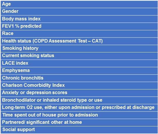
The hospitalist physician, after receiving verbal approval from the hospitalized COPD patient of his/her potential willingness to see an investigator for a clinical research study, was then seen by an investigator, and informed consent was obtained. Following this, an interview and review of medical records were performed to obtain demographic and disease variables. Variables (from interview or record review) included: demographics (age, gender), disease severity, all-cause and respiratory-related hospitalizations over the preceding year, outpatient treated respiratory exacerbations over the preceding year, functional status, co-morbidities, psychological status, treatment upon admission. COPD assessment test (CAT) (7), Charlson Comorbidity Index (CCI) (8) and LACE Index (9) were determined for all patients. We also measured the treating physician’s “gut feeling” of the likelihood of a 30-day readmission. The treating physician was blinded as to the specific variables we measured. (All variables tested are detailed in Appendix. Post-bronchodilator forced expiratory volume in one second (FEV1), forced vital capacity (FVC) and FEV1/FVC ratio were obtained from previous spirometry (within 3 years), if available. The patients without a historical spirometric diagnosis of COPD had spirometry before hospital discharge. Consented patients were then contacted at 30-days to determine whether they had readmissions and if so, for what cause.

General statistics are reported as means ± standard deviations (SD). Univariate logistic regression analyses were used to determine which of our tested variables predicted 30-day admission for exacerbation of COPD. Following this, multivariate forward logistic regression, incorporating variables that were predictive in univariate analyses, was utilized to determine which variables were predictive of 30-day hospitalization for COPD exacerbations.

Hospitalizations were analyzed as binary variables (yes-no). Based on the univariate analysis, two scoring systems were developed to predict readmission. The 2 scoring systems, each including three variables, significantly predicted 30-day readmissions.

The first scoring system (scoring system I) was as follows:

  1. MRC dyspnea. This score ranges from 0 (least) to 4 (greatest) dyspnea. Our scoring was dichotomized to 0 (MRC 0, 1, 3, or 3) or 1 (MRC 4: “too short of breath to leave the house or short of breath dressing/undressing.”
  2. Exacerbation history: Those with 1 or more hospitalizations for exacerbations in the preceding year were given a score of 1; those below this threshold had a score of 0.
  3. Chronic prednisone use prior to admission: Chronic prednisone use was defined as prednisone used on all or most days for at least three months prior to admission. Those meeting this criterion were given a score of 1, those without chronic prednisone use had a score of 0.

The second scoring system (scoring system II) was as follows:

  1. MRC dyspnea. This was identical to # 1 in the first scoring system.
  2. Exacerbation history: Those with 2 or more outpatient -treated exacerbations (some of these could result in hospitalization) in the preceding year were given a score of 1; those below this threshold had a score of 0.
  3. Chronic prednisone use prior to admission: This was identical to # 3 in the first scoring system.

Scores for each of the above scoring systems could, therefore, range from 0-3. The relationship between the above scores and 30-day hospital readmissions were evaluated using receiver operating characteristic (ROC) curves, which plot the true-positive rate (sensitivity) versus the false-positive rate (1-specificity).

A receiver operating characteristic (ROC) curve, plotting the true-positive rate (sensitivity) versus the false-positive rate (1-specificity) was used to characterize the relation. The ROC model was used to predict the likelihood of readmission for scoring system I and scoring system II.

Results

Of the 50 studied patients, 77% were female; mean age was 71 ± 11 years. The body mass index (BMI) was 29.65 + 9 kg/m2. Clinical characteristics of subjects are shown in Table 1.

Table 1. Clinical characteristics of studied subjects.

Sixty percent had Medical Research Council (MRC) dyspnea 3 or 4 (moderate to severe). Mean length of stay was 4.3 ± 3.2 days. Thirty-four percent lived alone at home.  

In our study, all patients readmitted within thirty days had respiratory exacerbations of COPD as principal diagnoses (i.e., the frequency of respiratory-related and all-cause 30-day readmissions was identical). Thirty-day readmission rate for exacerbation of COPD was 24%. Of the studied parameters, the ones that did not predict rehospitalization in univariate logistic regression analyses are shown in Table 2.

Table 2. Variables that did not predict 30-day readmission.

Variables that significantly predicted or tended to predict readmission included: 1) two or more clinical exacerbations (not necessarily resulting in hospitalization) in the previous year (OR 4.6, p= 0.04); 2) prednisone use (chronic or prior to admission) (OR 4.4, p< 0.04); 3) MRC = 4 (OR 2.7, p = 0.16); 4) one or more respiratory hospitalizations in the preceding year (OR 3.1, p = 0.08).

Using scoring system I, 16 patients had a score of 0; 16 had a score of 1, 14 patients had a score of 2, and 4 had a score of 3. Readmission rates for each of these categories were as follows: 13%, 19%, 29%, and 75%, respectively. Using the ROC model (Figure 1), odds ratios for readmission for- Score 0 versus 3 was 18; (2) odds ratios for readmission for score 1 versus 3 was 16 and (3) odds ratios for readmission for score 2 versus 3 was 6.7.

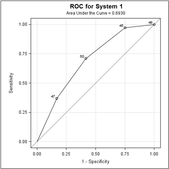
Figure 1. Receiver operating characteristic (ROC) curve for scoring system I, showing odds ratio for readmission for Score 0 versus 3, Score 1 versus 3 and Score 2 versus 3.

In scoring system II, 19 had a score of 0, 16 had a score of 1, 11 had a score of 2, and 4 had a score of 3. Readmission rates for each of these categories were as follows: 11%, 19%, 36%, and 75%, respectively.  Using the ROC model (Figure 2), odds ratios for readmission for- Score 0 versus 3 was 24; (2) Score 1 versus 3 was 15 and (3) Score 2 versus 3 was 4.5.

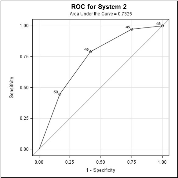
Figure 2. Receiver operating characteristic (ROC) curve for scoring system II, showing odds ratio for readmission for score 0 versus 3, score 1 versus 3 and score 2 versus 3.

In both scoring systems, the combined score of 3, with all 3 variables present, was associated with a high rate of readmission. The odds ratio was calculated for the clinical scores as it provides a valid effect measure and allows comparison of the clinical scores with regards to outcome, i.e. the readmission for COPD exacerbation, in a small study such as this.

The closer AUC is to 1, the better the predictive performance of the test, with the practical lower limit for the AUC of a predictive test being 0.5. In this study, scoring system I with an AUC of 0.69 (Figure 1) and scoring system II, with an AUC of 0.73 (Figure 2), indicate fair strength as predictors for COPD readmission.

Discussion

The purpose of our study was to create a simple scoring system that might predict 30-day readmissions in patients hospitalized with COPD exacerbations. Data regarding factors which predisposes to hospital readmissions within 30 days of discharge after hospitalization for acute exacerbations of COPD is variable and remains limited (4-6, 10,11). Our study aimed at identifying potential risk factors and evaluating probable predictors of hospital re-admission in COPD patients within a month of discharge.

In our study, three variables held promise in a scoring system used to predict re-hospitalization within 30 days: exacerbations (either clinically-treated or hospitalized), chronic prednisone use, and functional limitation from dyspnea. These three variables could be assessed within a few minutes from a review of the inpatient hospital record or from a brief interview.

Previous studies evaluating readmission risk factors in COPD up to one year have identified several variables. These include: a lower FEV1 (12- 16), reduced physical activity, functional limitation and poor health-related quality of life (2,4,17-19), need for self-care assistance, active/ passive smoking, long term supplemental O2-requirement (12,16-18), and presence of selected co-morbid conditions (20, 21).

More recent studies found low physical activity to be a significant factor (5,18). Minutes of physical activity per day in the first week following discharge was lower in those readmitted (42 + 14 minutes vs. 114 + 19 minutes, p = 0.02) (5). Ngyuen et al. (19) reported an 18% readmission rate in 4000 patients, with independent predictors of increased readmission including reduced activity, anemia, prior hospitalizations, longer lengths of stay, more comorbidities, receipt of a new oxygen prescription at discharge, use of the emergency department or observational stay before the readmission. In another retrospective study, multivariate analysis showed the following risk factors to be associated with early readmission within 30 days of discharge- male gender, history of heart failure, lung cancer, osteoporosis, and depression; no prior prescription of statin within 12 months of the index hospitalization and no prescription of short-acting bronchodilator, oral steroid and antibiotic on discharge; length of stay, <2 or >5 days and lack of follow-up visit after discharge (10). Another study found these variables to have a significant association with 30-day readmissions: age, diastolic blood pressure, COPD severity score, length of stay, pH, paCO2, FEV1< 50%, number of previous days until exacerbation (6). This study also found an increased mortality at 6 months and one year in patients readmitted within 30 days of discharge (6).

In our study, the most influential variable 30-day readmission was the history of two or more exacerbations in the preceding year (OR: 2.47, CI= 1.51-4.05, p< 0.001). This variable was also found in our study to be significantly associated with 30-day readmission, following discharge for a COPD exacerbation hospitalization.

Our study found steroid use (chronic or prior to admission) to be a significant predictor of COPD readmissions. Steroid use has been associated with a significantly increased risk of readmission in a few other studies (12,13,16,22). We hypothesize chronic prednisone use reflects instability and variability in the chronic respiratory disease or a recent exacerbation prior to the index hospitalization- hence its relatively strong relationship to re-hospitalization.

The second significant predictor in our study, exacerbations resulting in hospital admission in the preceding year, has been found to be a risk factor readmission in prior studies (6,12,13,23). Three admissions in the year preceding recruitment was found to increase risk for readmission for COPD exacerbation (12,13,23). Frequent exacerbations in the preceding year likely reflect the severity of disease in these patients. A retrospective study found no association between the number of previous hospital COPD admissions and readmission (24).

Our third significant predictor, the severity of dyspnea has also been reported in some studies to be an independent risk factor for hospital admission for an acute exacerbation of COPD. Kessler et al. (14) reported that COPD patients with a dyspnea of grade 3, 4 or 5 (defined as breathlessness with mild, minimal or limited exertion respectively), had a significant risk of hospitalization at one year but those with dyspnea of grade 2 did not. Patients with “severe dyspnea” have been found to be more likely to be readmitted to hospital in studies (15,18). Our study using the MRC rating for dyspnea and found patients with an MRC rating of 4, which is equal to the most severe grading of dyspnea in this scale. The severity of dyspnea by MRC dyspnea being a predictor for readmission in COPD indicates that the severity of the disease predisposes to exacerbations of COPD and consequent readmissions.

A systematic review of studies on risk factors for readmission for patients with COPD exacerbation found 3 predictive factors similar to our study, namely- previous hospital admission, dyspnea and oral corticosteroids (25). This review also identified other variables including use of LTOT, having low health status or poor health related quality of life and reduced routine physical activity as risk factors for admission and readmission for COPD exacerbation (25).

A scoring system similar to ours, using 3 the significant predictors of COPD readmission (chronic prednisone use, MRC dyspnea rating and prior exacerbations, either clinical or requiring hospitalizations) has not been studied in predicting the 30 day- readmission for COPD exacerbation. This scoring system was a fairly strong predictor of readmission for COPD and may serve as a useful tool in risk-stratifying patients and directing medical resources toward those patients most at risk for readmission. This is especially of relevance at the present time when hospitals will face financial penalties for 30-day readmissions for COPD.

The risk factors identified for COPD readmission in this study are not modifiable. However, if patients more at risk for readmissions can be identified based on these risk factors, more resources can be directed to these group of patients- such as closer outpatient follow-up, VNA services, inpatient and outpatient pulmonary rehabilitation, more gradual steroid taper and institution of anti-inflammatory therapy such as azithromycin.

One limiting factor of this study is the small number of patients. The scoring system generated by the study using the 3 identified predictors, though fairly predictive of readmissions for COPD exacerbations, cannot be used without corroboration. The validity of the scoring system using needs to be established in a larger group of patients. Based on the results of this study, we intend to assess these variables as part of a quality assurance study on a larger number of hospitalized COPD patients. We plan to attempt to refine the scoring system, if possible, with an emphasis on simplicity in assessing data, brevity in data collection and predictive power for 30-day and subsequent hospitalization.

Conclusions

A simple 3-point scoring system, incorporating three variables: 1) chronic prednisone use; 2) MRC dyspnea rating; and 3) prior exacerbations (either clinical or requiring hospitalizations) has a fairly high predictive value for 30 -day readmission due to COPD exacerbation. This can be easily assessed within a few minutes from a review of the inpatient hospital record or from a brief patient interview. It can serve as a useful tool in risk-stratifying patients and directing medical resources toward those patients most at risk for readmission. This scoring system using these three variables holds promise for future validation studies.

References

  1. Celli BR, Barnes PJ. Exacerbations of chronic obstructive pulmonary disease. Eur Respir J. 2007;29:1224-38. [CrossRef] [PubMed]
  2. Steer J, Gibson GJ, Bourke SC. Predicting outcomes following hospitalization for acute exacerbations of COPD. QJM. 2010;103:817-29. [CrossRef[ [PubMed]
  3. Johannesdottir SA. Hospitalization with acute exacerbation of chronic obstructive pulmonary disease and associated health resource utilization: a population-based Danish cohort study. J Med Econ. 2013;16:897-906. [CrossRef] [PubMed]
  4. Tan WC. Factors associated with outcomes of acute exacerbations of chronic obstructive pulmonary disease. COPD. 2004;1(2):225-47. [CrossRef] [PubMed]
  5. Sharif R, Parekh TM, Pierson KS, Kuo YF, Sharma G. Predictors of early readmission among patients 40 to 64 years of age hospitalized for chronic obstructive pulmonary disease. Ann Am Thorac Soc. 2014;11:685-94. [CrossRef] [PubMed]
  6. Guerrero M, Crisafulli E, Liapikou A, Huerta A, Gabarrus A, Chette A, Soler N, Torres A. Readmission for acute exacerbation within 30 days of discharge is associated with a subsequent increase in mortality risk in COPD patients: A long-term observational study. PLoS ONE. 2016;11:e0150737. [CrossRef] [PubMed]
  7. Jones PW, Harding G, Berry P, Wiklunf I, Chen WH, Kline Leady N. Development and first validation of the COPD assessment test. Eur Respir J. 2009;34:648-54. [CrossRef] [PubMed]
  8. Charlson M, Szatrowski TP, Peterson J, Gold J. Validation of a combined comorbidity index. J Clin Epidemiol. 1994:47:1245-51. [CrossRef] [PubMed]
  9. Walraven C, Dhalla IA, Bell C, Etchells E, Stiel IG, Zarnke K, Austin PC, Foster AJ. Derivation and validation of an Index to predict early death or unplanned readmission after discharge from hospital to community. CMAJ. 2010; 182: 551-7. [CrossRef] [PubMed]
  10. Garcia-Aymerich J, Monso E, Marrades RM, Escarrabill J, Felez MA, Sunyer J, Anto JM. Risk factors for hospitalization for a chronic obstructive pulmonary disease exacerbation. EFRAM study. Am J Respir Crit Care Med. 2001;164:1002-7. [CrossRef] [PubMed]
  11. Garcia-Aymerich J, Farrero E, Félez MA, Izquierdo J, Marrades RM, Antó JM. Risk factors of readmission to hospital for a COPD exacerbation: a prospective study. Thorax. 2003;58:100-5. [CrossRef] [PubMed]
  12. Gudmundsson G, Gislason T, Janson C, et al. Risk factors for rehospitalisation in COPD: role of health status, anxiety and depression. Eur Respir J. 2005;26:414–19. [CrossRef] [PubMed]
  13. Cao Z, Ong KC, Eng P, Tan WC, Ng TP. Frequent hospital readmissions for acute exacerbation of COPD and their associated factors. Respirology. 2006;11(2):188-95. [CrossRef] [PubMed]
  14. Lau AC, Yam LY, Poon E. Hospital re-admission in patients with acute exacerbation of chronic obstructive pulmonary disease. Respir Med. 2001;95:876-84. [CrossRef] [PubMed]
  15. Kessler R, Faller M, Fourgaut G, Mennecier B, Weitzenblum E. Predictive factors of hospitalization for acute exacerbation in a series of 64 patients with chronic obstructive pulmonary disease. Am J Respir Crit Care Med. 1999;159:158-64. [CrossRef] [PubMed]
  16. Wang Q, Bourbeau J. Outcomes and health-related quality of life following hospitalization for an acute exacerbation of COPD. Respirology. 2005;10:334-40. [CrossRef] [PubMed]
  17. Almargo P, Barriero B, DeEchaguen AO, Quintana S, Rodriguez CM, Heredia JL, Garau J. Risk factors for hospital re-admission in patients with chronic obstructive pulmonary disease. Respiration. 2006;73:311-7. [CrossRef] [PubMed]
  18. Chawla H, Bulathsinghala C, Tejada JP, Wakefield D, ZuWallack R. Physical activity as a predictor of thirty-day hospital re-admission after a discharge for a clinical exacerbation of COPD. Ann Am Thorac Soc. 2014;11:1203-9. [CrossRef] [PubMed]
  19. Ngyuen HQ, Chu L, Liu ILA, Lee JS, Suh D, Korotzer B, Yuen G, Desai S, Coleman KJ, Gould MK. Associations between physical activity and 30-day readmission risk in chronic obstructive pulmonary disease. Ann Am Thorac Soc. 2014;11(5): 695-705. [CrossRef] [PubMed]
  20. Kessler R, Faller M, Fourgaut G, Mennecier B, Weitzenblum E. Predictive factors of hospitalization for acute exacerbation in a series of 64 patients with Chronic Obstructive Pulmonary Disease. Am J Respir Crit Care Med. 1999;159:158–164. [CrossRef] [PubMed]
  21. Miravitlles M, Guerrero T, Mayordomo C, Sanchez-Agudo L, Nicolau F, Segu JL. Factors associated with increased risk of exacerbation and hospital admission in a cohort of ambulatory COPD patients: a multiple logistic regression analysis. Respiration. 2000;67:495–501. [CrossRef] [PubMed]
  22. Groenewegen KH, Schols AM, Wouters EF. Mortality and mortality-related factors after hospitalization for acute exacerbation of COPD. Chest. 2003; 124:459-67. [CrossRef] [PubMed]
  23. Connolly MJ, Lowe D, Anstey K, Hosker HSR, Pearson MG, Roberts CM. Admissions to hospital with exacerbations of chronic obstructive pulmonary disease: effect of age related factors and service organization. Thorax. 2006;61:843-8. [CrossRef] [PubMed]
  24. Pouw EM, Ten Velde GP, Croonen BH, Kester AD, Schols AM, Wouters EF. Early non-elective readmission for chronic obstructive pulmonary disease is associated with weight loss. Clin Nutr. 2000;19:95–99. [CrossRef] [PubMed]
  25. Bahadoori K, Fitzgerald JM. Risk factors of hospitalization and readmission of patients with COPD exacerbation-systematic review. Int J Chron Obstruct Pulmon Dis. 2007:2(3) 241-51. [PubMed]

Cite as: Yap V, Wilcox D, ZuWallack R, Datta D. Evaluating a scoring system for predicting thirty-day hospital readmissions for chronic obstructive pulmonary disease exacerbation. Southwest J Pulm Crit Care. 2018;16(6):350-9. doi: https://doi.org/10.13175/swjpcc054-18 PDF

Friday
Jun222018

Intralobar Bronchopulmonary Sequestration: A Case and Brief Review

Uddalak Majumdar, MD1 

Payal Sen, MD2

Akshay Sood, MD2

1Cleveland Clinic Foundation, Cleveland, OH USA

2Univeristy of New Mexico, Albuquerque, NM USA


Abstract

Objective: Bronchopulmonary sequestration is a rare congenital abnormality of the lower respiratory tract, seen mostly in children but often in adults. The term implies a mass of lung tissue that has no function and lacks normal communication with the rest of the tracheobronchial tree.

Case: A 40-year-old man presented with acute onset of left flank pain for 4 hours. He was born in Yemen and emigrated to the US in 1998; at that time, he had been tested for tuberculosis which was negative. In this admission, he met systemic inflammatory response (SIRS) criteria and had basilar crackles in the left lower lobe of the lung. CT scan revealed a cavitary lesion with air-fluid level in the left lower lobe airspace. There was systemic arterial blood supply to this region arising off the celiac axis. He was diagnosed with an infected intralobar bronchopulmonary sequestration and underwent video-assisted thoracoscopic wedge resection. On follow up 3 months later, he was doing well.

Discussion: Pulmonary sequestration is a rare congenital anomaly of a mass of lung tissue, which can have cystic changes and is a very important differential diagnosis of cavities in the lung. Confirmation of diagnosis is by visualization of a systemic vessel supplying sequestrated pulmonary, and this is accomplished by contrast-enhanced CT scan, MRI or invasive angiography. 

Conclusion: The delay in diagnosis in our patient was due to falling prey to anchoring and availability biases and chasing the diagnosis of tuberculosis in a patient from Yemen with a lower lobe cavitation.

Case

History of Present Illness: A 40-year-old man with a past medical history of atrial fibrillation presented to the hospital with acute onset of left flank pain for 4 hours, fevers and chills. The pain was sharp and stabbing, pleuritic, non-radiating, and was severe with an intensity of 10/10. He denied extraneous activity or trauma earlier in the day, denied substernal pain, cough, night sweats, weight loss or change in urinary habits. He was born in Yemen and emigrated to the US in 1998; at that time, he was tested for tuberculosis (TB) which was negative. He was known to have a cavitary lesion in left lower lobe since 2005, and had undergone extensive evaluation (imaging, sputum and PPD) which showed no form of tuberculosis. He denied taking prophylactic TB treatment. Annual PPD testing had always been negative.

The patient worked on a ship, which travelled in the Great Lakes on the US-Canada border. He was a current smoker with a 20-pack-year smoking history. He lived at home with his wife and children. There was no history of IV drug use, prior imprisonment or homelessness. He denied being in contact with anyone with TB while in Yemen. He was sexually active with his wife and had no other sexual partners. He denied history of sexually transmitted infections.

Physical Examination:

Vital Signs: Temp – 38.3 degrees Fahrenheit, Pulse- 111/minute, RR- 18/min, BP- 151/66 mm Hg. Spo2- 90 % on Room Air.

Basilar crackles and rhonchi in the left lower lobe of the lung. No cervical or inguinal lymphadenopathy. Rest of the physical exam was normal.

Significant Laboratory Findings:

WBC elevated at 15,500/mm3 with 65 percent Neutrophils.

Lactate - 1.1 mmol/dL

Radiography:

Chest x-ray was done while in the emergency department, which revealed left basilar sub-segmental atelectasis (Figure 1).

Fig.1. Chest x-ray showing left basilar sub-segmental atelectasis without focal consolidation, large pleural effusion or pneumothorax.

Initial CT scan of abdomen and pelvis was done to rule out renal/ureteral stone. It showed a left lower lobe airspace consolidation with bronchiectasis and bronchiolectasis and a cavitary lesion with air-fluid level (Figure 2). 

Figure 2. Representative images from the CT scan in lung windows showing left lower lobe airspace consolidation concerning for an acute on chronic process.

C-reactive protein and erythrocyte sedimentation rate were normal, CRP and ESR- normal; blood cultures revealed no growth; procalcitonin 0.4 ng/mL (normal <0.15); anti-nuclear antibody – negative; Aspergillus antigen – negative; urine Legionella antigen – negative; Streptococcus pneumoniae antigen – positive.

Sputum Gram stain and acid-fast bacilli culture/stain could not be obtained because the patient did not produce any sputum.

Subsequently CT chest with IV contrast was done which showed findings compatible with a pneumonia within a left lower lobe intrapulmonary sequestration. (Figure 3).

Figure 3. Representative images from the thoracic CT chest with IV contrast. The left lower lobe demonstrates a 69 x 83 mm heterogeneous fluid collection with multiple locules of air. There was systemic arterial blood supply to this region arising off the celiac axis (arrows).

The patient was diagnosed with an infected intralobar bronchopulmonary sequestration. He was treated initially with intravenous fluids and piperacillin-tazobactam. He underwent video-assisted thoracoscopic wedge resection of infected bronchopulmonary sequestration in left lower lobe and ligation of the systemic feeding vessels from the celiac artery. Pathologic examination revealed a fibrotic lung with areas of centrilobular emphysema, bronchiolectasis, mucus pooling and microscopic honeycomb changes. Findings also showed an elastic artery, with features most suggestive of intralobar sequestration. His symptoms completely resolved after his operation.

Discussion

Bronchopulmonary sequestration is a rare congenital abnormality of the lower respiratory tract, seen mostly in children but often in adults, like in our patient (1). In 1946, Pryce coined the term "pulmonary sequestration" to describe a disconnected bronchopulmonary mass or cyst with an anomalous arterial supply (2). The term implies a mass of lung tissue that has no function and lacks normal communication with the rest of the tracheobronchial tree. This mass of non-functional lung tissue receives blood supply from the systemic circulation (3). The exact etiology is unknown and is thought to be an embryologic process error in foregut budding (4), although some have indicated a non-congenital acquired process in intralobar sequestration.

Sequestration may be intra- or extralobar based on its relation with the normal lung lobes. An intralobar sequestration (ILS), like the name suggests, is located within a normal lobe, lacks its own visceral pleura (5) and also has aberrant connections to bronchi, and lung parenchyma, or even the gastrointestinal tract, and often presents with recurrent infections (6,7). Compared to ILS, an extralobar sequestration (ELS) is located outside the normal lung and has its own visceral pleura (8), with the rare occurrence of infectious complications (9). About 75% of BPS is intralobar while 25% is extralobar (10). Bronchopulmonary sequestration is often associated with other congenital abnormalities like congenital diaphragmatic hernia, vertebral anomalies, congenital heart disease, pulmonary hypoplasia, colonic duplication, and congenital pulmonary airway malformation (11). 

Clinically, pulmonary sequestration is latent until infection leads to symptoms (12). Symptoms, like that of any pathological lung condition depend on the type, size, and location of the lesion. Sepsis and extracardiac shunting are common complications of untreated sequestration. Hemoptysis can also be a presentation. The mechanism of pneumonia is post-obstructive and usually recurrence of pneumonia leads to diagnosis. Recurrent pneumonia especially in the lower lobes should always include intralobar sequestration in the differential diagnoses. But the pathophysiology of infection and/or hemoptysis when ILS is not connected to airway is a mystery. Sometimes there is a partial or anatomically abnormal connection to the tracheobronchial tree, which can lead to poor mucus clearance, plugging and recurrent infection.

The mainstay of diagnosis is pre-operative imaging and post-operative histopathology of the resected specimen. The pathognomonic imaging characteristic is systemic vascular supply of the affected area of the lung (intra or extra-lobar), which is seen in about 80% of CT scans. Recurrent infection can lead to cystic areas within the mass (clusters of “ring shadows” on X-ray) (13). The surrounding normal lung may have air trapping and show emphysematous changes. Radiologic signs of BPS are a spectrum and represent the chronic and recurrent inflammation of the sequestrated lung: recurrent focal airspace disease, a parenchymal mass, a cavitary consolidation or mass, cystic lesions, localized bronchiectasis or adjacent emphysema. Bronchoscopy has little role in the management of BPS, which needs to be kept in mind by clinicians investigating cystic lung lesions. Identifying the systemic feeding vessel also helps with surgical planning.  

Symptomatic patients are treated with surgical excision; surgery is curative and is associated with minimal morbidity (14). Surgery is urgent in patients with significant respiratory distress but may be an elective procedure in adults or older children with less symptoms (15, 16). 

For asymptomatic patients of any age, management depends on how ‘high risk’ they are considered for developing complications. High risk patients are those with large lesions occupying >20 percent of the hemithorax, bilateral or multifocal cysts, or those with pneumothorax. In these patients, surgical resection is preferred to observation (17). On the other hand, in asymptomatic patients without these high-risk characteristics, either elective surgical resection or conservative management with observation are reasonable options (18). 

Apart from surgery, even embolization of the anomalous arterial supply has been reported to result in a complete resolution of symptoms and imaging changes to a certain in some cases (19). Since identification of vascular supply during surgery may be difficult during surgery, presurgical embolization may reduce risk of vascular complications (19). Embolization also has a more important role in hemoptysis and heart failure from shunting.

Conclusions

  • Pulmonary sequestration is a rare congenital anomaly of a mass of lung tissue without a normal connection to the tracheobronchial tree and a systemic vascular supply.
  • Presentation in adults is due to complication of the mass, undiagnosed in childhood. 
  • Sequestrated lung can have cystic changes and is a very important differential diagnosis of the cavitation. 
  • Confirmation of diagnosis is by visualization of a systemic vessel supplying sequestrated pulmonary, and this is usually accomplished by contrast-enhanced CT scan, MRI or invasive angiography.

Teaching points

This is a case of adult presentation of congenital pulmonary malformation and represents a delay in diagnosis, even though the patient’s symptoms started 10 years ago. The delay was due to falling prey to anchoring and availability biases and chasing the diagnosis of TB ten years ago in a patient from Yemen with a lower lobe cavitation. 

The feeding vessel from the celiac axis can only be demonstrated via a contrast enhanced CT, and thus, when in doubt, we should always get angiography by contrast-enhanced-CT or MRI or by invasive angiography. Had it been thought of and done 10 years ago, the patient would’ve been diagnosed and treated earlier.

Disclosure Statement

Drs. Majumdar, Sen and Sood have no conflicts of interest or financial ties to disclose.

References

  1. Landing BH, Dixon LG. Congenital malformations and genetic disorders of the respiratory tract (larynx, trachea, bronchi, and lungs). Am Rev Respir Dis. 1979 Jul;120(1):151-85. [CrossRef] [PubMed]
  2. John PR, Beasley SW, Mayne V. Pulmonary sequestration and related congenital disorders. A clinico-radiological review of 41 cases. Pediatric radiology. Pediatr Radiol. 1989;20(1-2):4-9. [CrossRef] [PubMed]
  3. Van Raemdonck D, De Boeck K, Devlieger H, et al. Pulmonary sequestration: a comparison between pediatric and adult patients. Eur J Cardiothorac Surg. 2001 Apr;19(4):388-95. [CrossRef] [PubMed]
  4. Gezer S, Taştepe I, Sirmali M, Findik G, Türüt H, Kaya S, Karaoğlanoğlu N, Cetin G. Pulmonary sequestration: a single-institutional series composed of 27 cases. J Thorac Cardiovasc Surg. 2007 Apr;133(4):955-9. [CrossRef] [PubMed]
  5. Shanti CM, Klein MD. Cystic lung disease. Semin Pediatr Surg. 2008 Feb;17(1):2-8. [CrossRef] [PubMed]
  6. Stocker JT, Drake RM, Madewell JE. Cystic and congenital lung disease in the newborn. Perspect Pediatr Pathol. 1978;4:93-154. [PubMed]
  7. Schwartz MZ, Ramachandran P. Congenital malformations of the lung and mediastinum--a quarter century of experience from a single institution. J Pediatr Surg. 1997 Jan;32(1):44-7. [CrossRef] [PubMed]
  8. Abbey P, Das CJ, Pangtey GS, Seith A, Dutta R, Kumar A. Imaging in bronchopulmonary sequestration. Send to J Med Imaging Radiat Oncol. 2009 Feb;53(1):22-31. [CrossRef] [PubMed]
  9. Houda el M, Ahmed Z, Amine K, Amina BS, Raja F, Chiraz H. Antenatal diagnosis of extralobar pulmonary sequestration. Pan Afr Med J. 2014;19:54. [CrossRef] [PubMed]
  10. Frazier AA, Rosado de Christenson ML, Stocker JT, Templeton PA. Intralobar sequestration: radiologic-pathologic correlation. Radiographics. 1997 May-Jun;17(3):725-45. [CrossRef] [PubMed]
  11. Kravitz RM. Congenital malformations of the lung. Pediatr Clin North Am. 1994 Jun;41(3):453-72. [CrossRef] [PubMed]
  12. Hang JD, Guo QY, Chen CX, Chen LY. Imaging approach to the diagnosis of pulmonary sequestration. Acta Radiol. 1996 Nov;37(6):883-8. [CrossRef] [PubMed]
  13. Hernanz-Schulman M. Cysts and cystlike lesions of the lung. Radiol Clin North Am. 1993 May;31(3):631-49. [PubMed]
  14. Samuel M, Burge DM. Management of antenatally diagnosed pulmonary sequestration associated with congenital cystic adenomatoid malformation. Thorax. 1999 Aug;54(8):701-6. [CrossRef] [PubMed]
  15. Haller JA, Jr., Golladay ES, Pickard LR, Tepas JJ, 3rd, Shorter NA, Shermeta DW. Surgical management of lung bud anomalies: lobar emphysema, bronchogenic cyst, cystic adenomatoid malformation, and intralobar pulmonary sequestration. Ann Thorac Surg. 1979 Jul;28(1):33-43. [CrossRef] [PubMed]
  16. Al-Bassam A, Al-Rabeeah A, Al-Nassar S, Al-Mobaireek K, Al-Rawaf A, Banjer H, et al. Congenital cystic disease of the lung in infants and children (experience with 57 cases). Eur J Pediatr Surg. 1999 Dec;9(6):364-8. [CrossRef] [PubMed]
  17. Parikh DH, Rasiah SV. Congenital lung lesions: Postnatal management and outcome. Semin Pediatr Surg. 2015 Aug;24(4):160-7. [CrossRef] [PubMed]
  18. Singh R, Davenport M. The argument for operative approach to asymptomatic lung lesions. Semin Pediatr Surg. 2015 Aug;24(4):187-95. [CrossRef] [PubMed]
  19. Eber E. Adult outcome of congenital lower respiratory tract malformations. Swiss Med Wkly. 2006 Apr 15;136(15-16):233-40. [PubMed]

Cite as: Majumdar U, Sen P, Sood A. Intralobar bronchopulmonary sequestration: A case and brief review. Southwest J Pulm Crit Care. 2018;16(6):343-9. doi: https://doi.org/10.13175/swjpcc075-18 PDF