
Correct!
4. The nodule shows no evidence of enhancement of 4 post-contrast time points, suggesting a benign etiology
The solitary pulmonary nodule enhancement study shows that the nodule has not changed in size or morphology since presentation- no satellite nodules or cavitation is seen. The nodule shows no evidence of enhancement at any of the 4 post-contrast time points, which is a predictor that the nodule is benign. The value of solitary pulmonary nodule enhancement CT protocol lies in the ability to exclude malignancy when no enhancement (˃15 HU) is seen at any of the 4 post-contrast time points compared to the initial unenhanced imaging. Enhancement within a nodule, however, is non-specific and may occur with malignancies as well as benign lesions, such as hamartomas.).
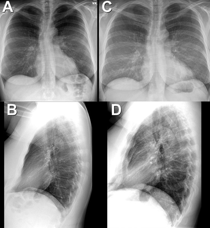
The patient did well over the next few years, with no recurrence of chest pain. She underwent follow up chest radiography (Figure 7) 4 years after her initial presentation.

Figure 7. Frontal (A) and lateral (B) chest radiography performed 4 years after initial presentation compared to presentation (C and D). To view Figure 7 in a separate, enlarged window click here.
Which of the following statements regarding this chest radiograph is most accurate? (Click on the correct answer to be directed to the 10th of 17pages)