
Correct!
4. Frontal and lateral chest radiography shows the left lower lobe nodule has increased in size
The nodule is difficult to perceive, best appreciated in the retrocardiac region on the frontal chest radiograph but appears larger compared to presentation chest radiography (Figure 1). No peribronchial or mediastinal lymph node enlargement is present, no other nodules are seen, and no pleural disease is evident.
The patient continued to do well from a cardiac perspective, with her eosinophilic endomyocarditis considered resolved. No major medical issues over the 9 years following her initial presentation. She remained under the care of family practice, with several screening chest radiographs (Figures 6 and 7) ordered, which were interpreted as largely unchanged from the initial presentation. Nine years after her initial presentation, the patient presented to the Emergency Room with complaints, of heavy, sharp chest pain of 10 days’ duration, which prompted repeat frontal chest radiography (Figure 8).

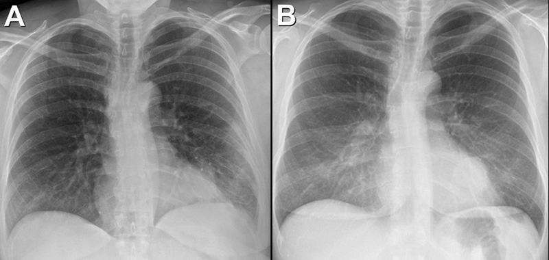
Figure 8. Frontal chest radiography performed 9 years after initial presentation (A) shows patchy subpleural left lung opacity (arrowheads), new from the most recent chest radiograph performed 4 years earlier (B). The left lower lobe nodule (arrow) is evident in both studies. To view Figure 8 in a separate, enlarged window click here.
Which of the following statements regarding this chest radiograph is most accurate? (Click on the correct answer to be directed to the 11th of 17pages)