
Correct!
5. More than one of the above
Given the apparent solid, vascularized appearance at chest CT, a neoplasm is a likely consideration and hence further staging of the lesion is warranted and 18FDG-PET would prove useful in this regard. Enhanced MR may also be beneficial as the superior contrast resolution of MR makes it the preferred choice for imaging of the local extent of chest wall neoplasms. Obtaining a tissue diagnosis is also appropriate, and percutaneous transthoracic biopsy is an ideal choice because the lung will not be entered to sample this lesion and hence the risk of complication is low, and this procedure also allows a large core biopsy to be performed for diagnosis. Thoracic surgical consultation is also appropriate.
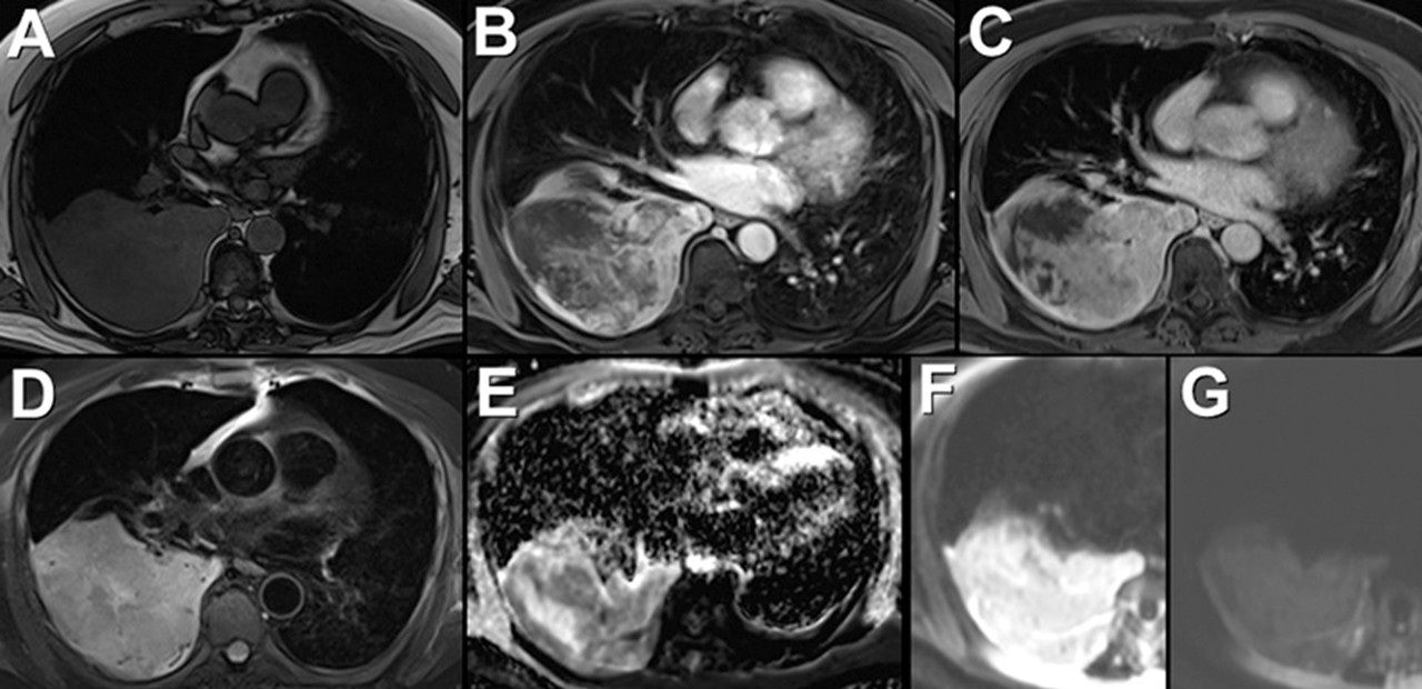
Thoracic surgical consultation was obtained and recommended that the patient undergo unenhanced and enhanced chest MR (Figure 6) for further evaluation of the right posterior chest mass.


Figure 6. Left: Unenhanced (A, D, E, and F) and enhanced (B and C) chest MR. To view the Figure 6 in a separate enlarged window click here. Right: video of enhanced chest MRI.
Regarding this examination, which of the following is correct? (click on the correct answer to be directed to the ninth of 11 pages)