
Correct!
5. The chest MR adds no new or unique information to the CT findings
The chest MR shows that the right-sided chest mass has extensive contact with the right posterior chest wall soft tissues and the lesion enhances substantially, confirming its vascularized nature. There is no evidence of fluid within the lesion and the lesion appears solitary- there is no other evidence of either lung nodules or enhancing nodules within the thoracic soft tissues. While non-specific, the MR assessment, using diffusion-weighted imaging, suggests that the lesion is cellular and therefore likely neoplastic.
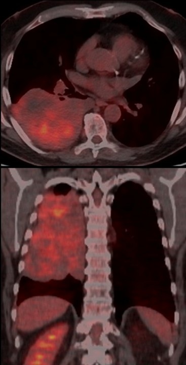
The patient then underwent 18FDG-PET scanning (Figure 7) for further evaluation of the right posterior chest mass.

Figure 7. 18FDG-PET. To view Figure 7 in a separate, enlarged window click here.
Regarding this examination, which of the following is correct? (click on the correct answer to be directed to the tenth of 11 pages)