
Correct!
1. Enhanced thoracic CT
In general chest CT is the most appropriate next step for evaluation of a mass detected at chest radiography. Chest MR may be employed for some lesions arising from the chest wall or osseous lesions, but when used for chest wall lesions, MR is typically the examination of choice when the lesion is palpable, raising suspicion for lymphadenopathy or sarcoma. 18FDG-PET scanning may play a role for chest masses as well, although typically after the lesion has been further characterized with chest CT. 99mTAA- ventilation-perfusion scintigraphy is employed for the evaluation of suspected acute or chronic thromboembolic disease or to assess relative lung perfusion as part of preoperative planning; neither are considerations in this circumstance. Finally, 68Ga-citrate scanning has been used for evaluation of diffuse lung diseases, especially infections, but is rarely employed for such now, and is not used for the assessment of a mass detected at chest radiography.
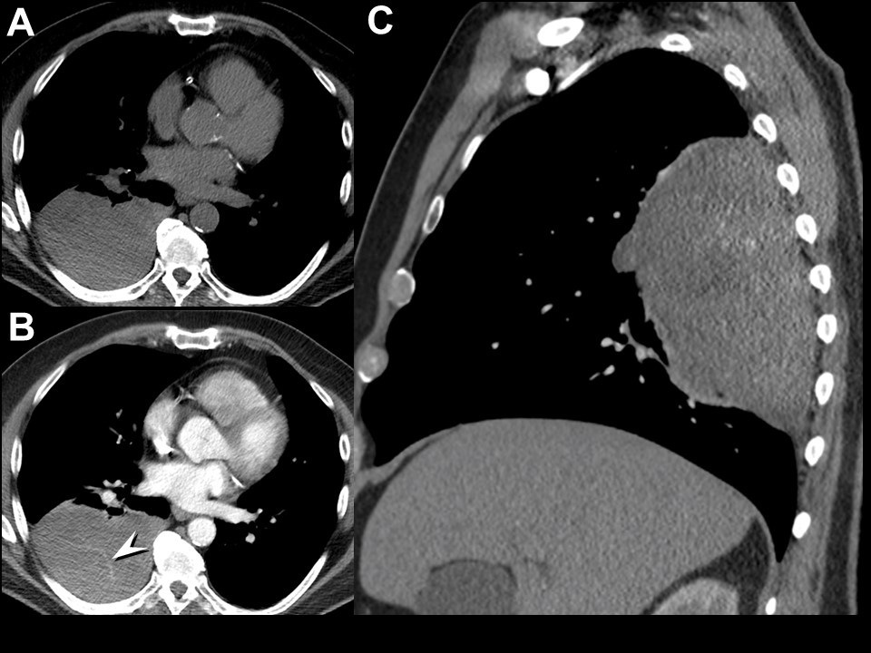
The patient underwent unenhanced and enhanced chest CT (Figure 4) for further evaluation of the chest radiographic abnormality.


Figure 4. Left (A-C): Representative views of unenhanced chest CT in soft tissue windows. To view figure 4A-C in a separate, enlarged window click here. Right: Video of enhanced chest CT. To view the video in a separate, enlarged window click here.
Regarding this examination, which of the following is correct? (click on the correct answer to be directed to the sixth of 11 pages)