
Correct!
5. The chest CT shows an oblong soft tissue mass in the right lower thorax.
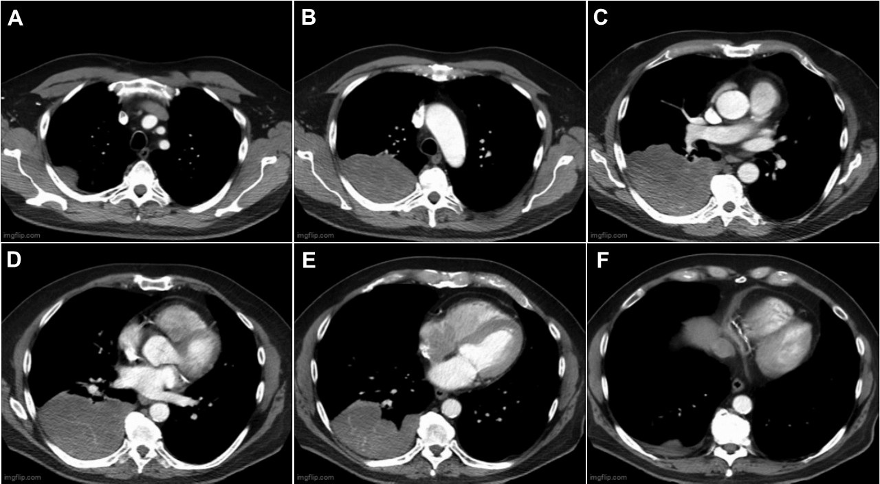
The chest CT was performed with both unenhanced (Figure 4) and enhanced (Figure 5) technique, which allows assessment for potential enhancement of the chest lesion; enhancement would suggest the lesion is vascularized and hence not simple pleural fluid (attenuation coefficients before and after intravenous contrast administration increased from 26 HU to 37 HU).


Figure 5. Left (A-F): representative images of enhanced CT in soft tissue windows. To view Figure 5 a separate, elaraged window click here. Right: Video of enhanced chest CT in soft tissue windows.
The lesion shows extensive, non-dependent contact with the right posterior chest wall and some faint internal linear hyperattenuation, suggesting vascularity, is visible. Given the “incomplete border” sign at chest radiography, and the extensive chest wall contact, the lesion very likely arises from extraparenchymal soft tissues and not the lung, and the lesion does not show cavitation. The lesion does not arise from the posterior mediastinum, nor does it exhibit osseous reaction or destruction.
Which of the following is not a correct choice for the differential diagnosis for the posterior right chest lesion identified at chest radiography and CT? (click on the correct answer to be directed to the seventh of 11 pages)