
Correct!
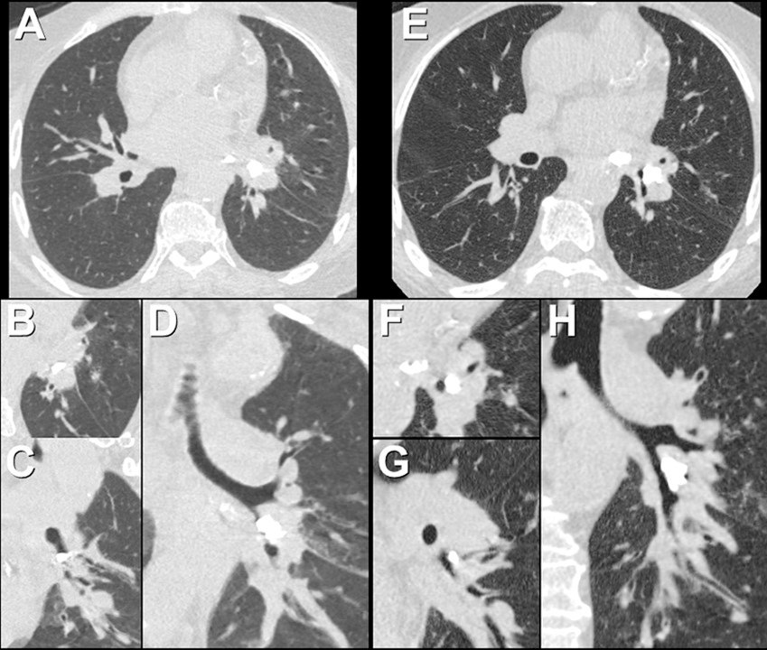
3. Unenhanced chest CT shows persistent lingular and left lower lobe consolidation and bronchiolitis, and a new left pleural effusion
Unenhanced chest CT shows the left lower lobe broncholith appears to completely obstruct the left lower lobe bronchus, and the lingular and left lower lobe consolidation and bronchiolitis persist. See Figure 10, which is comparison of the repeat examination, Figure 9, with the CT performed at the time of broncholithiasis diagnosis, Figure 6.

Figure 10. Comparison of the repeat chest CT (A-D) with the CT performed at the time of diagnosis of broncholithiasis (E-H). (A) Axial unenhanced image with oblique axial (B), oblique sagittal (C), and oblique coronal (D) reformatted images. To view Figure 10 in a separate enlarged window, click here.
A new left pleural effusion is now evident. No new right-sided pulmonary opacities are seen.
Based on the information thus far, which of the following is the most appropriate course of action? (Click on the correct answer to be directed to the twelth and final page)