
Correct!
3. Repeat thoracic surgery consultation
Given that the lingular and left lower lobe pneumonia has not resolved, a repeat thoracic surgery consultation for treatment of the broncholith is in order. Neither chest MR nor 18FDG – PET scan plays a role for the management of this patient.
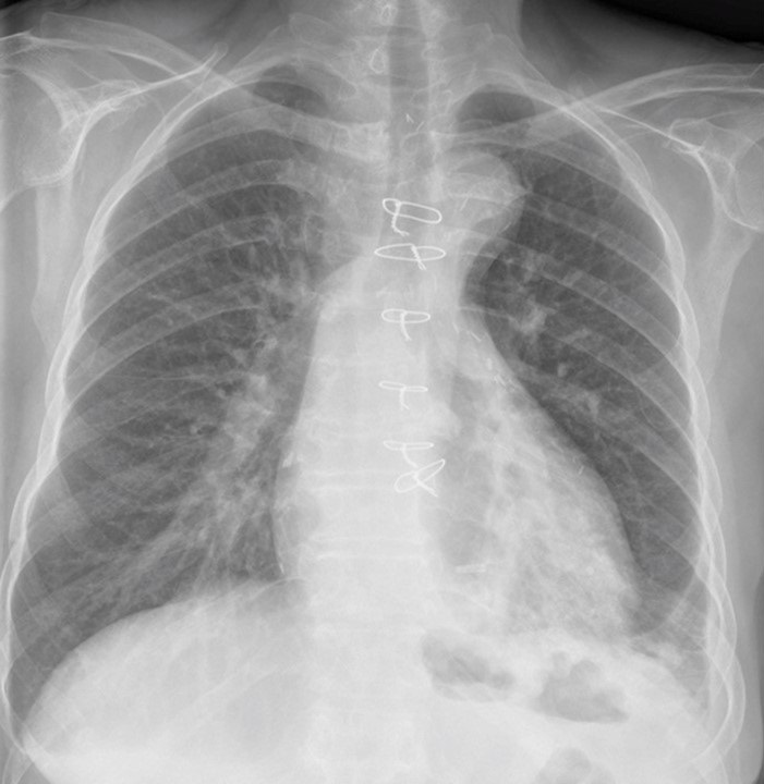
Over the course of the ensuing year, the patient presented to the Emergency Room with various complaints, including syncope, urinary tract infection and two episodes of left lower lobe pneumonia. The thoracic surgeon was again reluctant to intervene. Just over a year and a half after broncholithiasis was diagnosed, the patient again presented with left lower lobe pneumonia (Figure 11).

Figure 11. Frontal chest radiography one-and-a-half years after broncholithiasis was diagnosed, when the patient presented to the Emergency Room with cough and fever, shows left lower lobe consolidation consistent with pneumonia. To view Figure 11 in a separate, enlarged window, click here.
The patient underwent repeat bronchoscopy (Figure 12).

Figure 12. Repeat bronchoscopy one-and-a-half years after initial diagnosis of broncholithiasis, when the patient was diagnosed with recurrent left lower lobe pneumonia, again shows a large broncholith obstructing much of the left lower lobe bronchus.
The left lower lobe was again obstructed by a broncholith, but using saline dilation, the broncholith was bypassed and the anteromedial basilar, lateral basilar, and posterior basilar subsegments were visualized, although the superior segment of the left lower lobe could not be seen. The broncholith seemed to be loose in the airway, hence the patient was intubated with an 8.5mm endotracheal tube and, using a gastrointestinal grasping forceps, the broncholith was grasped and gently retracted and removed enbloc. Inspection of the left mainstem bronchus revealed some minor bleeding and clot obstructing the left lower lobe, which was removed. The carina between the left lower lobe and superior segment was intact and the left lower lobe was widely patent. The patient tolerated the procedure well and has had not suffered recurrent pneumonia since.
Diagnosis: Broncholithiasis with recurrent pneumonia
References