
Correct!
2. Frontal and lateral chest radiography shows a focal, poorly defined right base opacity
Frontal and lateral chest radiograph shows a focal, somewhat oblong and poorly defined right base opacity, seen on the frontal projection, but not well visualized on the lateral projection (Figure 2).
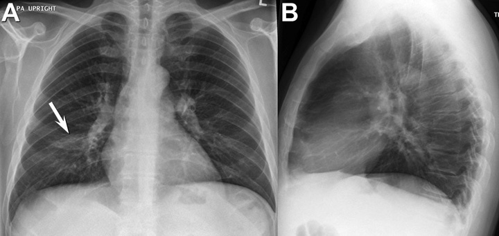
Figure 2. Frontal (A) and lateral (B) chest radiography shows a poorly defined, faintly nodular opacity at the right base (arrow), primarily seen on the frontal projection.
No evidence of peribronchial or mediastinal lymph node enlargement is seen, no are linear or reticular abnormalities. The heart size appears normal.
The patient underwent abdominal cross sectional imaging to address his complaint of weight loss and jaundice, which disclosed abnormalities of the right kidney and liver (Figure 3), with cholelithiasis and possibly some thickening of the extrahepatic bile ducts.
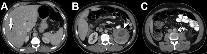
Figure 3. Top A-C: Representative images from axial enhanced abdominal CT showing heterogeneous wedge-shaped low attenuation in the liver (arrow), enlargement of the left kidney with a low attenuation lesion anteriorly (arrowhead) and diminished left renal enhancement, and some mild left periaortic thickening (double arrowheads). Bottom: video of abdominal CT scan.
Which of the following would be most useful for the evaluation of this patient? (Click on the correct answer to proceed to the third of seven pages)