Krystal Chan, MD
Bilal Jalil, MD
Department of Internal Medicine
University of New Mexico School of Medicine
Albuquerque, NM USA
A 48-year-old man with a history of hypertension, intravenous drug abuse, hepatitis C, and cirrhosis presented with 1 day of melena and hematemesis. While in the Emergency Department, the patient was witnessed to have approximately 700 mL of hematemesis with tachycardia and hypotension. The patient was admitted to the Medical Intensive Care Unit for hypotension secondary to acute blood loss. He was found to have a decreased hemoglobin, elevated international normalized ratio (INR), and sinus tachycardia. A bedside echocardiogram was performed.
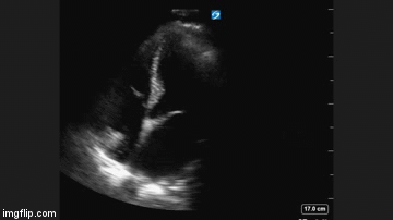
Figure 1. Apical four chamber view of the heart.
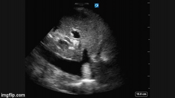
Figure 2. Longitudinal view of the inferior vena cava entering into the right atrium.
What is the best explanation for the echocardiographic findings shown above? (Click on the correct answer for an explanation and discussion)
Cite as: Chan K, Jalil B. Ultrasound for critical care physicians: now my heart is still somewhat full. Southwest J Pulm Crit Care. 2016;12(6):236-9. doi: http://dx.doi.org/10.13175/swjpcc054-16 PDF