
Correct!
5. All of the above
All of the listed differential diagnoses could produce the patient’s eosinophilia and could be consistent with her clinical presentation. Testing for HIV, Lyme disease, coccidioidomycosis, West Nile virus, EBV, Parvovirus 19, and CMV were all negative, as was testing for toxoplasmosis, strongyloidiasis, and filariasis, and Trichinella. Ova and parasite testing was negative. On the 5th hospital day, her troponin began to trend down (0.124 → 0.221 → 0.424 → 0.570 → 0.630, → 0. 447, ng/mL (normal, ≤0.01 ng/mL). The patient was discharged with cardiology, pulmonary, and oncology follow up scheduled.
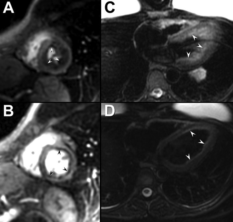
The patient’s chest pain resolved, and her eosinophilia decreased, and her corticosteroid therapy was tapered. Testing for genetic derangements associated with hypereosinophilia syndrome was negative. The patient underwent follow up cardiac MR (Figure 5) which showed decreased cardiac edema.

Figure 5. Cardiac MR performed 3 months after initial presentation following high-dose corticosteroid therapy shows persistent circumferential first-pass perfusion defects on the short axis first-pass / perfusion sequence (arrowheads, A= initial presentation, B= 3 month follow up examination), but improved T2-hyperintense subendocardial inflammation on the 4-chamber fat saturation T2-weighted sequence (arrowheads, C= initial presentation, D= 3 month follow up examination. To view figure 5 in a separate, enlarged window click here.
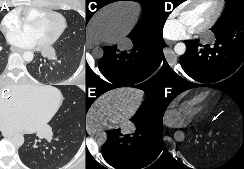
The patient also underwent repeat enhanced chest CT 6 months after the initial presentation (Figure 6) for further evaluation of the nodule discovered at presentation chest CT.

Figure 6. Unenhanced and enhanced chest CT performed according to a solitary pulmonary nodule enhancement protocol shows a stable medal left lower lobe pulmonary nodule (28 mm, A= presentation, B= 6 month follow up) without clear enhancement (C= unenhanced, D= 1 minute, E= 4 minutes, F= subtraction image. To view Figure 6 in a separate, enlarged image click here.
Note how the nodule appears similar to background lung on the subtraction image (arrow, F), indicating no internal enhancement.
Regarding this examination, which of the following is correct? (Click on the correct answer to be directed to the 8th of 17 pages)