
Correct!
1. CT pulmonary angiography
Given the history of chest pain and elevated D-dimer levels, CT pulmonary angiography may prove useful. Efforts directed at a tissue diagnosis at this point are premature. 18FDG-PET scan is potentially useful for staging lung cancer and evaluation of pulmonary nodules and thus may ultimately be of benefit for this patient but typically adds value after cross sectional imaging characterization. Enhanced chest CT is preferable to MR [with or without intravenous] for suspected lung parenchymal abnormalities.
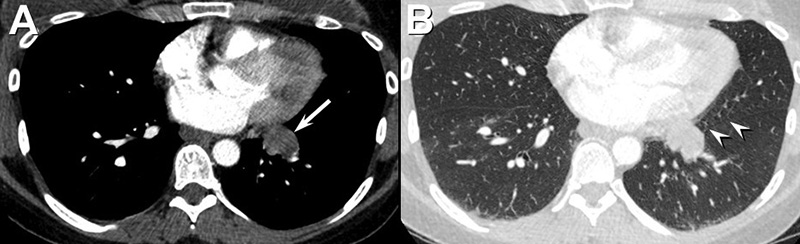
The patient underwent CT pulmonary angiography (Figure 3).


Figure 3. Axial enhanced CT pulmonary angiography (A, soft tissue, B, lung windows). To view figure 3 in a separate, enlarged window click here. Lower: Video of the pulmonary angiography in lung windows.
Which of the following statements regarding the CT pulmonary angiogram is most accurate? (Click on the correct answer to be directed to the 4th of 17 pages)