
Correct!
1. Enhanced chest CT
Given the normal D-dimer levels, CT pulmonary angiography is not required, although some form of enhanced chest CT may be of benefit given the known lung nodule. Enhanced chest CT is preferred over MR for the evaluation of lung pathology. 18FDG-PET scan is potentially useful for staging lung cancer and evaluation of pulmonary nodules but typically adds its greatest value after cross sectional imaging characterization. A tissue diagnosis may be required for the nodule, but further characterization with cross sectional imaging is required because the nodule is rather poorly characterized at chest radiography alone, although it does appear enlarged.
The patient underwent CT pulmonary angiography (Figure 9).


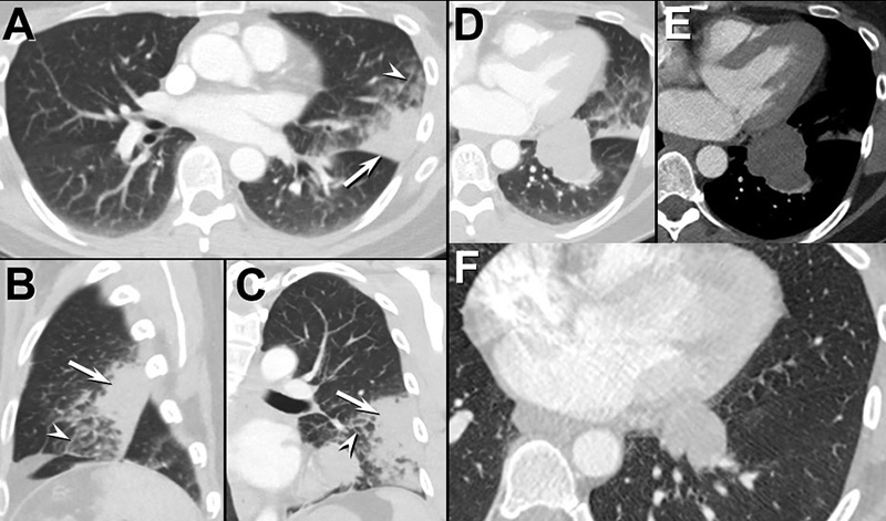
Figure 9. Left: Axial enhanced CT pulmonary angiography (A, axial, B, sagittal, C, coronal) shows pleural-based / subpleural lingual consolidation (arrows) corresponding to the chest radiograph. Smoothly thickened interlobular septae (arrowheads) are present. The appearance is consistent with pneumonia. D and E, Axial lung (D) and soft tissue (E) windows show the non-calcified left lower lobe lesion The chest CT 9 years earlier (F). To view Figure 9 in a separate, enlarged window click here. Right: video of CT pulmonary angiography in lung windows. To view the video in a separate, enlarged window click here.
Which of the following statements regarding the CT pulmonary angiogram is most accurate? (Click on the correct answer to be directed to the 13th of 17pages)