
Correct!
5. All of the above
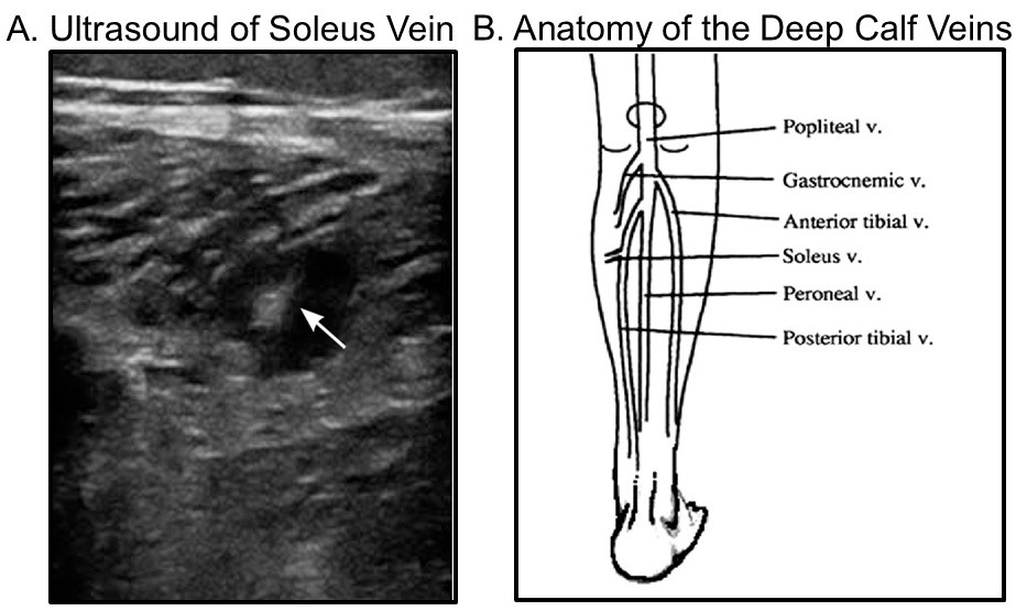
The lower extremity venous ultrasound was performed and shown in Figure 1A.

Figure 1. A (left): Ultrasound of the right soleus vein showing a venous thrombosis. B (right): Anatomy of the deep calf veins. To view Figure 1 in a separate, enlarged window click here.
Should the patient be anticoagulated? (Click on the correct answer to be directed to the fourth of six pages)