
Correct!
2. Frontal chest radiography shows bronchovascular thickening and new focal lung opacity
Frontal and lateral chest radiography shows new bronchovascular thickening and patchy new opacity overlying the heart on the lateral project [difficult to localize on the frontal projection], raising the possibility of pneumonia. The chest radiographic findings are subtle, and appreciation of the abnormalities is facilitated by direct comparison of the current study with the previous examination (Figure 5).

Figure 5. Comparison of lateral chest radiograph at presentation in the Emergency Room with complaints of productive cough (A) and the lateral chest radiograph 2 years earlier. The comparison highlights the interval development of bronchovascular thickening (arrowheads) and patchy, localized opacity overlying the heart, suggesting either right middle lobe or lingular pneumonia. To view Figure 5 in a separate enlarged window, click here.
No pleural abnormality is seen, and no new discrete nodules are apparent. No evidence of mediastinal lymph node enlargement is present.
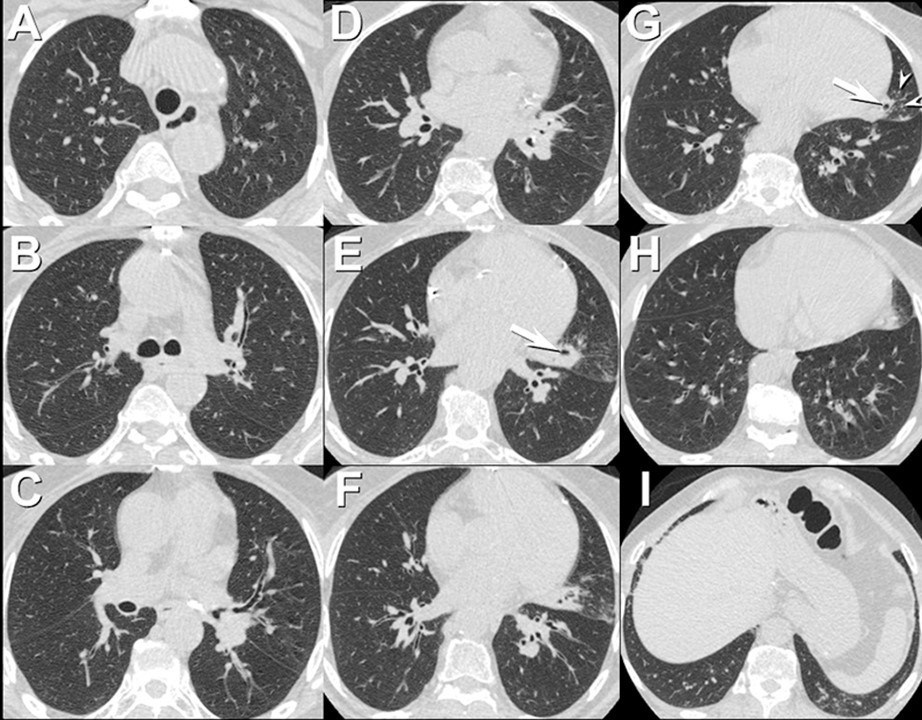
Nasal swabs for respiratory syncytial virus (RSV) were positive. Chest CT (Figure 6) was performed.

Figure 6. Representative images from axial unenhanced chest CT in lung windows. To view Figure 6 in a separate enlarged window, click here.
Which of the following represents an appropriate interpretation for this examination? (Click on the correct answer to be directed to the seventh of 12 pages)