
Correct!
5. More than one of the above
Given the patient’s immunosuppression, a heightened surveillance posture seems appropriate, and cross-sectional imaging of the chest, abdomen, and pelvis, particularly considering fever and possible hypotension, is reasonable. Chest MRI is not appropriate as CT will provide more effective pulmonary evaluation. 18FDG – PET scan is premature at this point, and is usually reserved for directed indications, such as cancer staging or nodule evaluation.
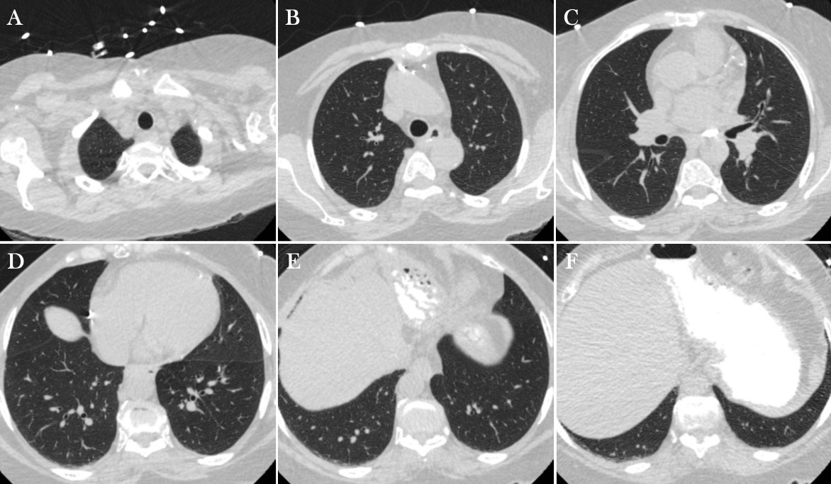
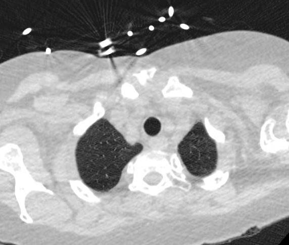
The patient underwent unenhanced chest CT (Figure 3) as well as CT of the abdomen and pelvis.


Figure 3. Left: Representative images from chest CT in lung windows. To view Figure 3 in a separate enlarged window, click here. To view a video of Figure 3, click here. Right: Video of chest CT.
Which of the following represents an appropriate interpretation for this examination? (Click on the correct answer to be directed to the fifth of 12 pages)