
Correct!
5. All of the above
The patient appears to be ill but does not seem to show signs of septic physiology. While he is at high risk for volume depletion, his exam is not suggestive of intravascular hypovolemia. He also is demonstrating evidence of hypercalcemia of undetermined etiology, which would merit IV hydration. Given the history of immunosuppression, a thorough workup for infection should be undertaken, including obtaining blood cultures and more advanced imaging to include chest and abdomen given his previous known chest abnormalities and a history of recent nausea and vomiting.
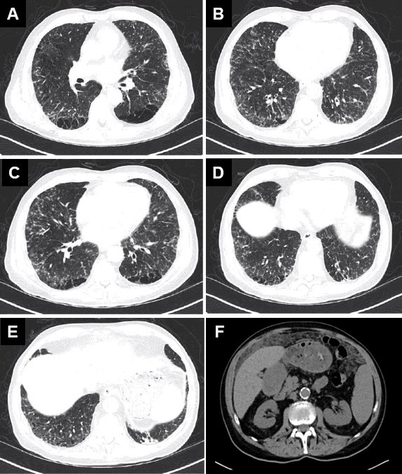
A CT scan of the chest, abdomen, and pelvis was performed (Figure 3).


Figure 3. Left: Representative images from admission CT chest (A-E) and abdomen (F). Right: Video of CT scan in lung windows. To view Figure 3 in a separate, enlarged window click here. To view a video of the CT scan click here.
What would be the next most appropriate step? (Click on the correct answer to be directed to the fourth of seven pages)