Correct!
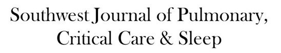
4. Repeat unenhanced chest CT shows multifocal ground-glass opacity in a somewhat different distribution than the prior two chest CTs
Unenhanced chest CT performed 18 weeks following initial presentation shows areas of bilateral, right-greater-than-left upper lobe predominant mild ground-glass opacity in a somewhat different distribution than either of the two previous chest CTs (Figures 2 and 4), but again without clear fibrotic features. There is mild architectural distortion best seen in the right upper lobe, but no clear bronchiolectasis is evident. No new pleural abnormality is seen. No new pulmonary nodules are evident, and no substantial new consolidation is seen.).
Given all the information presented thus far, which of the following represents the most appropriate next step for the patient’s management? (Click on the correct answer to be directed to the eleventh and final page)