Correct!
1. Unenhanced chest CT shows a small nodule
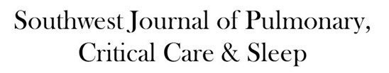
A small nodule is barely detectable in the medial left lower lobe. Some tubular opacity extending distally from this nodule suggests an association with a subsegmental airway. The nodule is retrospectively visible on the two previous chest CTs (Figure 8). The remainder of the enhanced chest appears normal.

Figure 8. Axial focused unenhanced CT images performed at presentation (Present) show a small nodule in the medial left basal initially overlooked. Images immediately cranial (A) and caudal (C) to the lesion (arrow, B) do not show an abnormality; the nodule (arrow) is visible on one image only. D-F: Axial focused unenhanced images less than one year following presentation again show the lesion visible on one image only (arrow, E) the nodule merges indistinctly with adjacent vessels. G-I: Axial focused enhanced CT 6 years following presentation and bilateral adrenalectomy, and following recurrence of symptoms suggesting Cushing syndrome, shows an enlarging medial left lower lobe nodule (arrows) at the site of the previously overlooked lesion on the two previous CTs. The nodule has a branching configuration (arrow in H), with tubular opacities distal to the lesion (arrowheads in I), suggesting an airway association.
Given all the information presented thus far, which of the following represents the most appropriate next step for the patient’s management? (Click on the correct answer to be directed to the eleventh of twelve pages)