Correct!
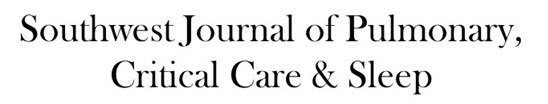
2. Enhanced chest CT shows a mass related to the chest wall
The enhanced chest CT sows that the lung parenchyma appears normal. There is no bronchiectasis and lung attenuation is homogeneous- there is no mosaic perfusion. No peribronchial or mediastinal lymph node enlargement is present. A homogeneous solid mass is present in the left posterior interior thorax.
Based on the appearance on the chest CT Which of the following represents an appropriate interpretation for this examination? (Click on the correct answer to be directed to the eighth of 11 pages)