
Medical Image of the Month: Diaphragmatic Eventration
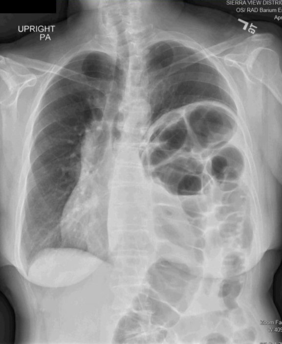
Figure 1. An upright PA chest radiograph demonstrates marked elevation of the left hemidiaphragm with associated superior migration of the gas-filled colon and mild mediastinal shift towards the right.
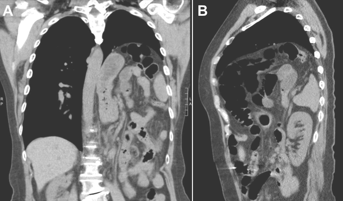
Figure 2. A: frontal. B: sagittal. A non-contrasted reconstruction of the chest demonstrates marked elevation of the left hemidiaphragm with associated superior migration of the abdominal viscera along with preservation of the integrity of the hemidiaphragm. These findings are consistent with a left hemidiaphragm eventration.
Clinical Presentation: A 66-year-old woman presented with a three-year history of progressive postprandial dyspnea and left-sided abdominal pain. Physical exam revealed normal vital signs and bowels sounds over left lung fields on auscultation. Laboratory work revealed a mild normocytic anemia. Imaging demonstrated marked left hemidiaphragm elevation with ipsilateral lung parenchyma volume loss and atelectasis along with a mild contralateral mediastinal shift. A sniff test was consistent with left hemidiaphragm paralysis.
The patient underwent a left video-assisted thoracoscopy, and the left hemidiaphragm was noted to be so thin that the abdominal organs could be visualize through it. The central tendon of the left hemidiaphragm was extremely attenuated and larger than normal. The left hemidiaphragm muscle fibers were noted to be situated around the periphery and not providing any significant tension. The redundant left hemidiaphragm central tendon was excised, and the patient was discharged without symptoms one week later.
Discussion: Eventration of a hemidiaphragm is a rare condition where there is non-paralytic weakening and thinning of a hemidiaphragm resulting in elevation of the hemidiaphragm with retained attachments to the costal margins (1). An eventration usually results from a congenital failure of the fetal diaphragm to muscularized. It is usually unilateral, occurs more on the right than the left, affects the anteromedial portion of the hemidiaphragm, occurs more often in women, and is found after the age of 60 in the adult population. A total eventration of a hemidiaphragm may be indistinguishable from diaphragmatic paralysis and result in a false-positive sniff test – as in this case. When symptomatic, it can pose a diagnostic challenge as it may be confused with a traumatic diaphragmatic rupture in the right clinical setting. Asymptomatic adults do not require treatment.
Leslie Littlefield MD and Mohamed Fayed MD
Department of Pulmonary and Critical Care
University of California San Francisco Fresno
Fresno, CA USA
Reference
- Black MC, Joubert K, Seese L, et al. Innovative and Contemporary Interventions of Diaphragmatic Disorders. J Thorac Imaging. 2019;34(4):236-247. [CrossRef] [PubMed]
Cite as: Littlefield L, Fayed M. Medical image of the month: diaphragmatic eventration. Southwest J Pulm Crit Care. 2020;21(1):9-10. doi: https://doi.org/10.13175/swjpcc036-20 PDF
 Post a Comment
Reader Comments