
Medical Image of the Month: Viral Pneumonias
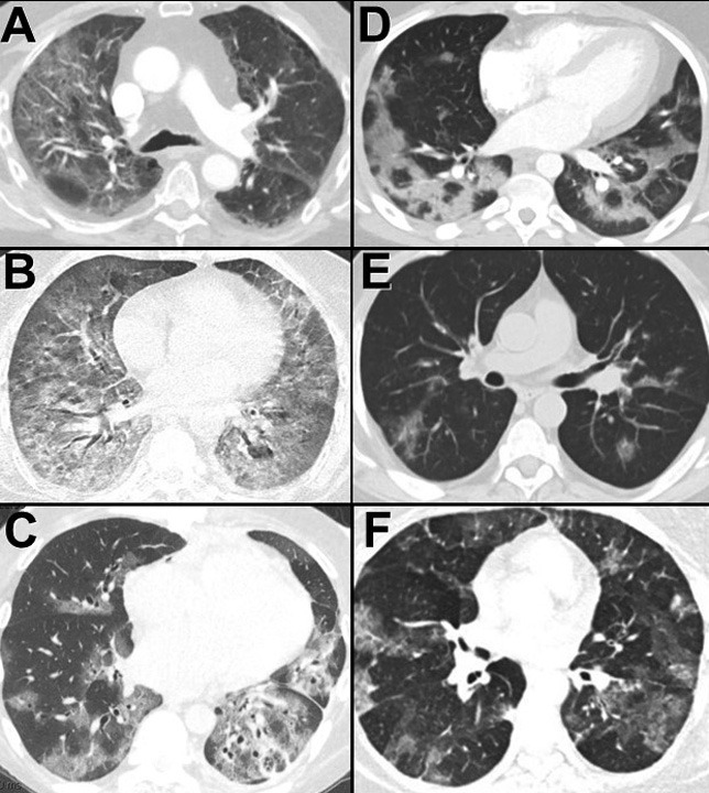
Figure 1. Pulmonary viral infection spectrum on thoracic CT scan in lung windows: A= Coronavirus NL63; B= Adenovirus; C= Influenza AH1 2009; D= COVID-19; E= Coronavirus HKU1; F= Influenza AH1 2009.
Numerous viruses, including the corona, influenza and adenoviruses can cause lower respiratory tract infection in adults (1). Viral pneumonia in adults can be classified into two clinical groups: so-called atypical pneumonia in otherwise healthy hosts and viral pneumonia in immunocompromised hosts. Until the COVID-19 pandemic, influenza virus types A and B caused most cases of viral pneumonia in immunocompetent adults. Immunocompromised hosts are susceptible to pneumonias caused by a wide variety of viruses including cytomegalovirus, herpesviruses, measles virus, and adenovirus. The CT imaging findings consist mainly of patchy or diffuse ground-glass opacity, with or without consolidation, and reticular areas of increased opacity, are variable and overlapping. The imaging findings in COVID-19 pneumonia are generally not distinctive compared to other viral pneumonias, including other coronaviruses such as SARS and MERS (2). A recent study systematically reviewed the longitudinal changes of CT findings in COVID-19 pneumonia. The results suggested that the lung abnormalities increase quickly after the onset of symptoms, peak around 6-11 days, and are followed by persistence of the findings.
Bacterial pneumonias may also take multiple forms and are sometimes difficult to radiographically separate from viral pneumonia (3). However, the presence of ground-glass opacities alone is unusual for a bacterial pulmonary infection. Rather, bacterial infections commonly present as areas of consolidation with air bronchogram formation, centrilobular nodules (often with branching configurations) and airway thickening.
Michael B. Gotway MD
Department of Radiology
Mayo Clinic Arizona
Scottsdale, AZ USA
References
- Kim EA, Lee KS, Primack SL, et al. Viral pneumonias in adults: radiologic and pathologic findings. Radiographics. 2002 Oct;22 Spec No:S137-49. [CrossRef] [PubMed]
- Wang Y, Dong C, Hu Y, Li C, Ren Q, Zhang X, Shi H, Zhou M. Temporal Changes of CT Findings in 90 Patients with COVID-19 Pneumonia: A Longitudinal Study. Radiology. 2020 Mar 19:200843. [CrossRef] [PubMed]
- Panse PM, Jokerst CE, Gotway MB. May 2020 Imaging Case of the Month: Still Another Emerging Cause for Infiltrative Lung Abnormalities. Southwest J Pulm Crit Care. 2020. May 1. (in press). [CrossRef]
Cite as: Gotway MB. Medical image of the month: viral pnuemonias. Southwest J Pulm Crit Care. 2022;20(5):163-4. doi: https://doi.org/10.13175/swjpcc028-20 PDF
 Post a Comment
Reader Comments