
Medical Image of the Week: Granulation Tissue
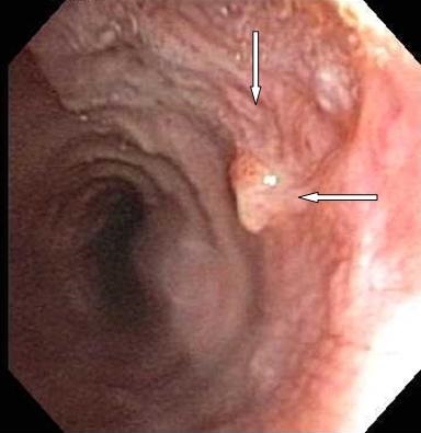
Figure 1. Subglottic space showing the presence of granulation tissue (arrows).
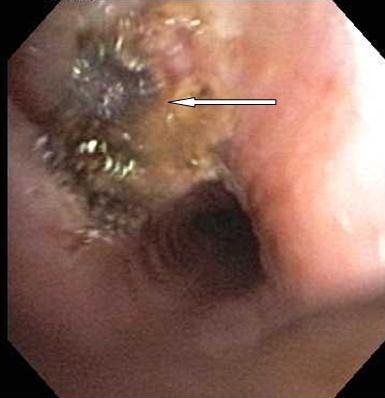
Figure 2. Argon Plasma Coagulation of the granulation tissue
A 57 year old woman presented with a tickling sensation in the back of throat and intermittent bleeding from the healing stoma one month after decannulation of her tracheostomy tube. On bronchoscopy a granuloma with surrounding granulation tissue was present in the subglottic space (Figure 1). Argon plasma coagulation (APC) was performed to cauterize the granulation tissue (Figure 2).
Formation of granulation tissue after tracheostomy is a common complication which can result in tracheal stenosis. APC and electrocautery using flexible bronchoscopy has been shown to safely and effectively remove the granulation tissue.
Aarthi Ganesh, MBBS and James Knepler, MD
Pulmonary, Allergy, Critical Care, & Sleep Medicine
University of Arizona
Tucson, AZ
Reference
- Epstein SK. Late complications of tracheostomy. Respir Care. 2005;50(4):542-9. [PubMed]
Reference as: Ganesh A, Knepler J. Medical image of the week: granulation tissue. Southwest J Pulm Crit Care. 2014;8(3):192-3. doi: http://dx.doi.org/10.13175/swjpcc029-14 PDF
 Post a Comment
Reader Comments