
Correct!
2. Large right pleural effusion
All the choices can cause an increased density on the x-ray in the right lower chest (Figure 2).
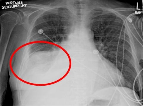
Figure 2. Admission portable chest x-ray showing the abnormality in the right lower chest (red circle).
There is a homogenous, dense haze over the right lower chest without any evidence of air bronchograms as usually seen with a pneumonia. Given the clinical situation of a fluid-overloaded patient, this makes right pleural effusion the most likely diagnosis. This was confirmed by ultrasound (Figure 3).
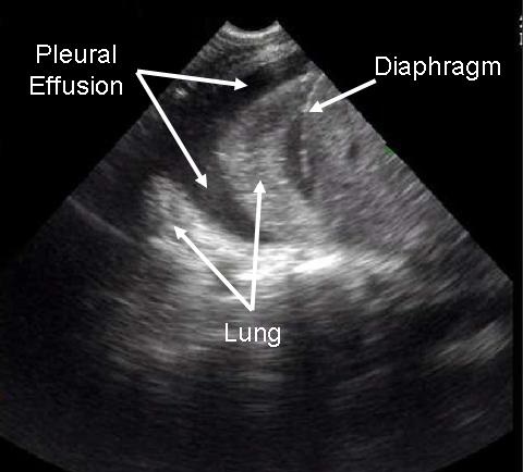
Figure 3. Right pleural view ultrasound showing pleural effusion.
Which of the following should be done next? (Click on the correct answer to move to the next panel)