
Correct!
1. Mixed obstructive and restrictive lung disease with moderately reduced DLco.
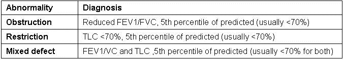
Although there are several protocols for interpreting pulmonary function testing, most are similar and the American Thoracic Society/European Thoracic Society guidelines are perhaps the most commonly used (1). The types of ventilatory defects are summarized in table 2.
Table 2. Summary of types of ventilatory defects and their diagnoses (1).
A thoracic CT was performed (Figure 1).
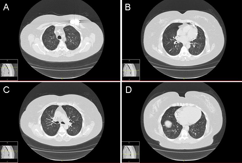
Figure 1. Panels A-D: Representative lung windows from the thoracic CT scan. Lower panel: movie of the lung windows from the thoracic CT scan.
Which of the following is the best interpretation of the thoracic CT scan? (Click on the correct answer to move to the next panel)