
Correct!
4. Serial chest x-rays
The patient has a history of lung cancer and new or changing back pain should raise a suspicion of metastatic disease, even 12 years after her thoracotomy. Plain x-rays necessitate a 1 cm diameter mass and 50% bone mineral loss at minimum for detection. Up to 40% of lesions will be unidentified by X-rays (1). The other choices are all reasonable for detecting metastatic disease (2). In this case a thoracic CT/PET scan was performed which showed a lesion in T5 (Figure 1, Panel A) which was biopsied (Figure 1, Panel B).
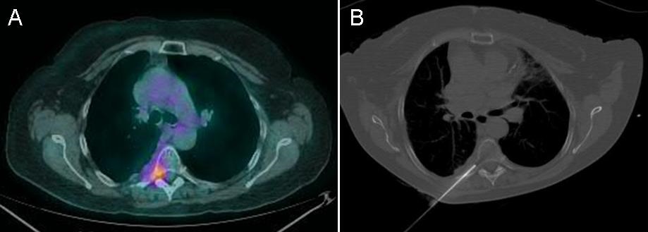
Figure 1. Panel A: CT/PET scan showing increased glucose metabolism in T5. Panel B: biopsy of lesion.
The biopsy showed adenocarcinoma. Immunochemical staining for thyroid transcription factor-1 (TTF-1) and napsin A were positive consistent with metastatic adenocarcinoma of the lung. Epidermal growth factor receptor (EGFR) and anaplastic lymphoma kinase (ALK) were negative. Radiation therapy was recommended.
In addition to the radiation therapy which of the following is the best choice for chemotherapy? (click on correct answer to move to next panel)