
Correct!
3. Massive pulmonary embolus
The CT angiogram shows a massive pulmonary embolus as evidenced by pulmonary artery intraluminal filling defects. This is seen in both the lung and soft tissue windows (Figure 3).
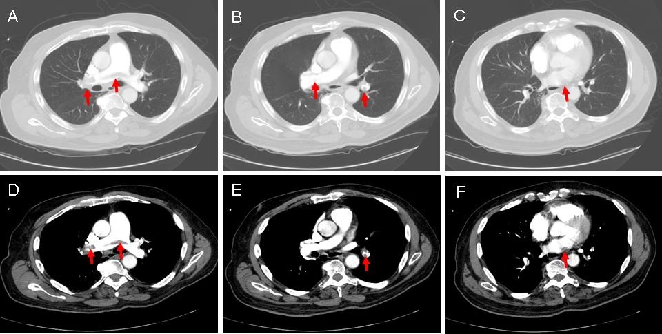
Figure 3. Intraluminal filling defects (red arrows).
The diagnosis is not surprising. A patient who becomes dyspneic, tachycardic and hypoxic post-operatively with a normal chest x-ray is at high risk for pulmonary embolism.
Which of the following is indicated at this time?