
Correct!
Pulmonary function testing is indicated to assess the severity of her lung disease, especially since her chest x-ray is normal. Pulmonary CT scanning is indicated to further define her chest disease. Rheumatologic evaluation is indicated to assess the activity of her rheumatoid arthritis. Repeat of the open lung biopsy would be overly aggressive at this juncture. Her pulmonary function testing revealed moderate restrictive disease with a moderate decrease in DLco.
Spirometry:
FVC 62% of predicted
FEV1 69% of predicted
FEV1/FVC 86%
FEF 25-75 89% of predicted
TLC 63% of predicted
DLco 47% of predicted
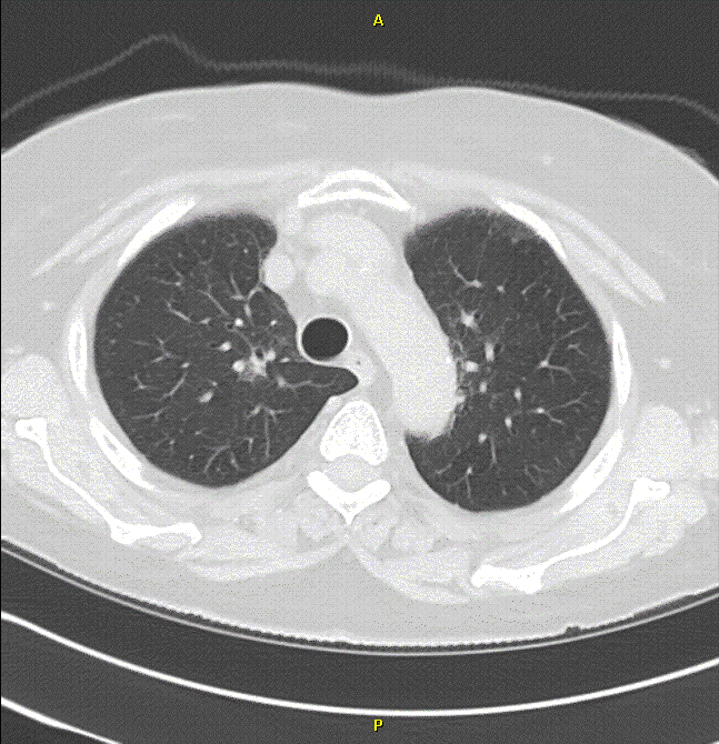
CT scanning was interpreted as showing scattered areas of ground glass opacities and bronchial wall thickening (Figure 1).
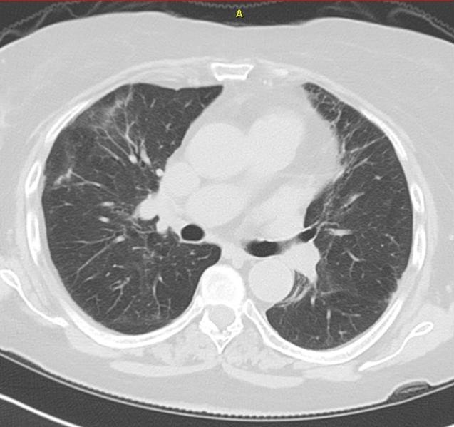
Figure 1. A representative lung window from thoracic CT scanning. (Click here for a movie of the CT scan)
Rheumatologic evaluation revealed the following:
Rheumatoid factor (RF): negative
Cyclic citrullinated peptide antibody (CCP): negative
Erythrocyte sedimentation rate: 3 mm/hr
Hand Films: no destructive changes
What is the next most logical step in her evaluation?