
Correct!
4. 1 and 3
The chest x-ray suggested an enlarging right apical bullae (Figure 2) which was confirmed on chest CT scan.
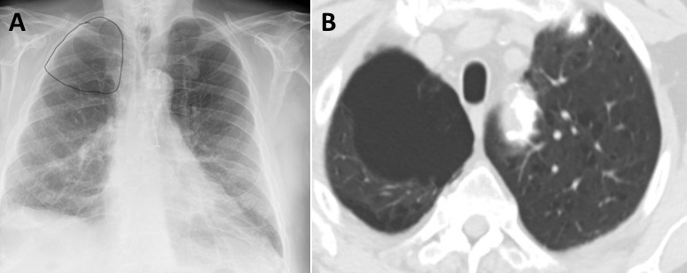
Figure 2. Panel A: Chest x-ray showing right lung bullae (circled). Panel B: Confirmation of enlarging bulla on CT scan.
It is very appropriate to admit an 80-year-old complaining of shortness of breath and hypoxic to the hospital often to the ICU. Theophylline is of dubious value in treating an exacerbation of COPD (1,2). Bronchodilators, antibiotics and corticosteroids have been shown to be of value (2).
He was admitted to the ICU and improved. After a short course of antibiotics, his prednisone was tapered and he was treated with an inhaled beta agonist on an as needed basis.
He did well until about 15 months later. He was admitted to a hospital in Vancouver with a small amount of hemoptysis. A representative image from a thoracic CT scan is shown in Figure 3.
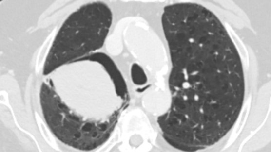
Figure 3. Representative image from thoracic CT in lung windows.
He did not recall the diagnosis although it sounded like “I see Tacoma”. He became concerned when his doctors discussed another thoracic operation and he came back to Phoenix for a second opinion.
Which of the following is the most likely diagnosis? (click on the correct answer to be directed to the third of five pages)