Correct!
5. Mycetoma
The CT scan shows a large mass-like opacity inside the previous identified bulla with a rim of air surrounding the mass, an air crescent sign. The most likely diagnosis from the CT findings was thought to be a mycetoma or Aspergillus fungus ball inside the bulla. The air crescent sign of a mycetoma, also referred to as the Monad sign, is seen in an immunocompetent host with preexisting cystic or cavitary lung disease, usually from tuberculosis or sarcoidosis (3). The fungal ball or mycetoma develops within a preexisting lung cavity and may exhibit gravity dependence. The mycetoma is composed of fungal hyphae, mucus, and cellular debris. Mycetomas can cause hemoptysis. The treatment options include surgical resection, bronchial artery embolization, and antifungal agents. The air crescent sign is not specific for Aspergillus infection and can be seen in other conditions, such as cavitating neoplasm, intracavitary clot, and Wegener’s granulomatous pneumonitis with angiitis (GPA) (3). Note that p-ANCA, c-ANCA and MPO and PR3 antibodies were negative.
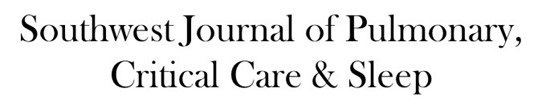
A bronchoscopy was performed (Figure 4).

Figure 4. Bronchoscopy showing small amount of blood coming from the superior subsegment of the right lower lobe.
Washings from the RLL showed no growth and cytology was negative.
What should be done next? (click on the correct answer to be directed to the fourth of five pages)