Correct!
5. Frontal chest radiography shows multifocal peribronchial consolidation
Frontal chest radiography shows multifocal, bilateral, somewhat mid and upper lung predominant peribronchovascular thickening and peribronchial consolidation. No pleural effusion is seen. The heart size is not markedly abnormal, and mediastinal lymphadenopathy is not readily apparent.
The patient underwent CT pulmonary angiography (Figure 2).
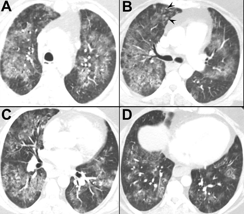
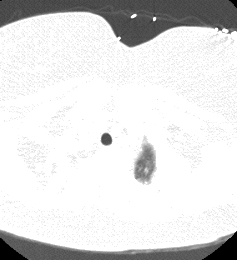
Figure 2. Left (A-D): Representative images from axial enhanced CT pulmonary angiography in lung windows. Right: Video of pulmonary angiography.
Which of the following represents an appropriate interpretation for this examination? (Click on the correct answer to be directed to the third of 11 pages)