Correct!
1. Frontal chest radiography shows normal findings
The patient underwent enhanced chest CT (Figure 2) given her complaint of shortness of breath.
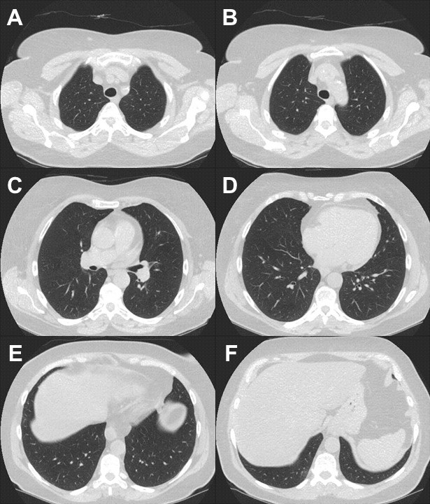
Figure 2. Left: representative images from axial unenhanced chest CT. Right: video of unenhanced chest CT.
Which of the following represents an appropriate interpretation for this examination? (Click on the correct answer to be directed to the second of twelve pages)