
Correct!
1. Active Tuberculosis (TB)
The patient has mediastinal lymphadenopathy and all of the above answer choices are potential etiologies for intrathoracic lymphadenopathy in patients with HIV. Given the patients history of latent TB, cough, duration of symptoms, and history of homelessness the best answer is active tuberculosis.
The differential for intrathoracic lymphadenopathy in patients with HIV includes: mycobacterial disease, nontuberculous mycobacterium disease, bacterial/fungal infection, sarcoidosis, and malignancy (1). Intrathoracic lymphadenopathy in HIV-infected patients is a common finding, in a study by Jasmer et al. (2) 35% of HIV-infected patients undergoing chest CT were found to have intrathoracic lymphadenopathy. Opportunistic infections were present in 60% of patients having intrathoracic lymphadenopathy on chest CT (2). Differentiating tuberculosis from lymphoma can be difficult but there are a number of clinical and radiographic predictors (Table 1).
Table 1. Predictors of mycobacterial disease.
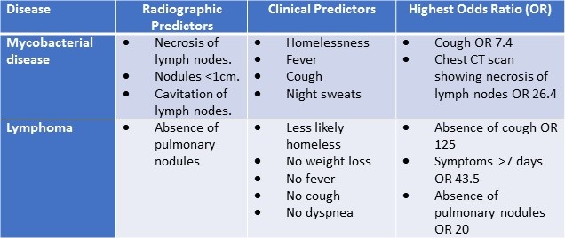
Radiographic and clinical predictors of mycobacterial disease include: homelessness, fever, cough, night sweats, necrosis of lymph nodes, nodules <1cm, cavitation (2). Factors associated with the highest likelihood of tuberculosis and/or nontuberculous mycobacterium include: cough OR 7.4 and chest CT scan showing necrosis of lymph nodes OR 26.4 (2). Radiographic and clinical predictors of lymphoma include: less likely homeless, no weight loss, no fever, absence of cough OR 125, no dyspnea, symptoms >7 days OR 43.5, absence of pulmonary nodules OR 20 (2).
The patient was subsequently started on broad spectrum antibiotics while awaiting cultures and placed in a negative pressure room.
Given the patients lymphadenopathy and history what would be your next step to establish a diagnosis? (Click on the correct answer to be directed to the fourth of six pages)