
Correct!
5. More than one of the above
The “best” answer may be comparison to prior studies, which is always the first step when evaluating an imaging abnormality. Nevertheless, high-resolution chest CT is also a reasonable choice, because the imaging findings is compelling and probably merit investigation in this symptomatic patient regardless of the chronicity as documented by comparison with prior chest radiographs. Even bronchoscopy with transbronchial biopsy is not wholly unreasonable as an interstitial abnormality associated with respiratory symptoms is present, but a tissue diagnosis is probably still premature at this point. 18FDG-PET scanning in not appropriate, however. Typically, 18FDG-PET scanning is used to investigate solitary pulmonary nodules or to stage patients with malignancies, and is not typically employed for the evaluation of interstitial lung abnormalities.
Prior chest radiography from 5 years earlier (Figure 2) was located.
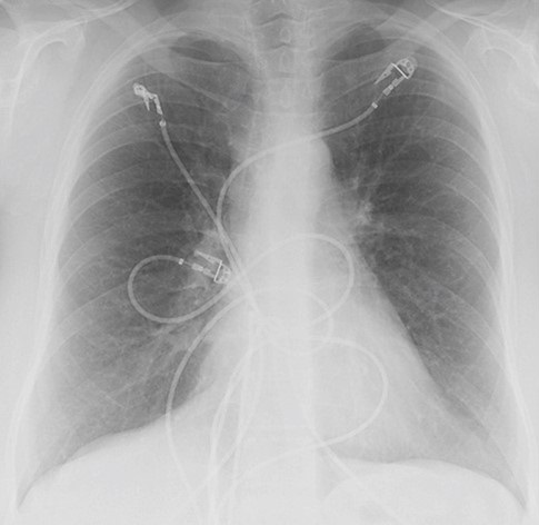
Figure 2. A comparison frontal chest radiograph performed 5 years earlier.
Which of the following represents the most accurate assessmentof the current and prior chest radiographic findings? (Click on the correct answer to proceed to the fourth of ten pages)