
Correct
5. Thoracic CT shows small pulmonary nodules
Thoracic CT shows multiple small pulmonary nodules, many of which are closely related to the pleura, or are frankly subpleural, and represent perifissural nodal tissue, reflecting intrapulmonary lymph nodes (Figures 2-5). A more focal opacity is present within the right suprahilar region, in the posterior portion of the apical segment of the right upper lobe; this focus has a different morphology than the other numerous, bilateral subcentimeter pulmonary nodules. There is no evidence of bronchiectasis or basilar fibrotic changes, and no ground-glass opacity is seen. None of the small nodules show evidence of cavitation.
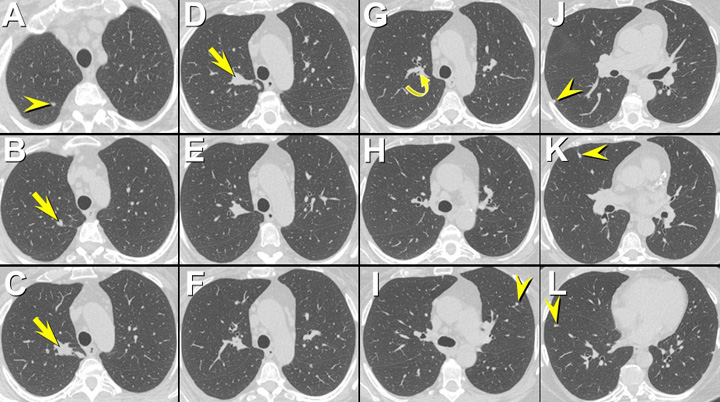
Figure 2 Unenhanced axial thoracic CT displayed in lung windows shows multiple, bilateral, peripheral and frankly subpleural, subcentimeter circumscribed nodules (arrowheads) bilaterally. Many of these small nodular opacities are very closely related to the costal and fissural pleural surfaces and are consistent with perifissural nodules, which typically reflect benign intrapulmonary lymph nodes. No evidence of cavitation within the nodules is seen, and no ground-glass opacity, bronchiectasis, or fibrotic changes are present. A focal opacity (arrows) with a somewhat tubular configuration (arrow in B) is present. This lesion is closely related to right upper lobe apical subsegmental airways. A small endobronchial lesion (curved arrow) may be present within a right upper lobe apical subsegmental airway.
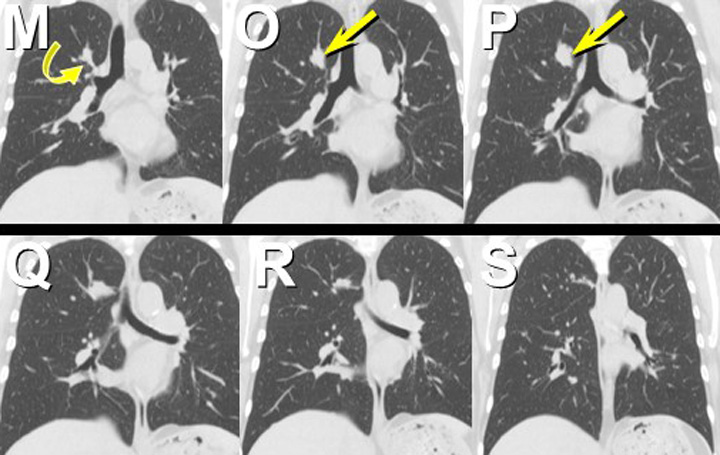
Figure 3. Unenhanced thoracic CT reformatted in the coronal plane focused on the opacity within in the right upper lobe apical segment shows the somewhat nodular character (arrow) of the lesion. A possible relationship between the lesion and right upper lobe apical subsegmental airways (curved arrow) is evident in this projection.
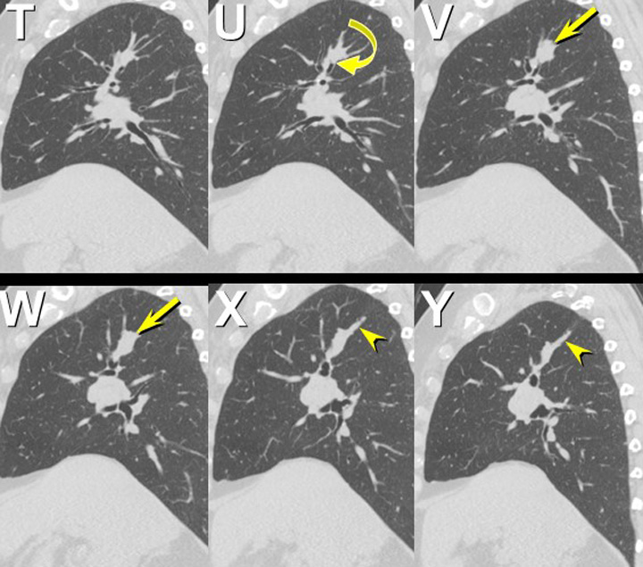
Figure 3. Unenhanced thoracic CT reformatted in the coronal plane focused on the opacity within in the right upper lobe apical segment shows the somewhat nodular character (arrow) of the lesion. A possible relationship between the lesion and right upper lobe apical subsegmental airways (curved arrow) is evident in this projection.
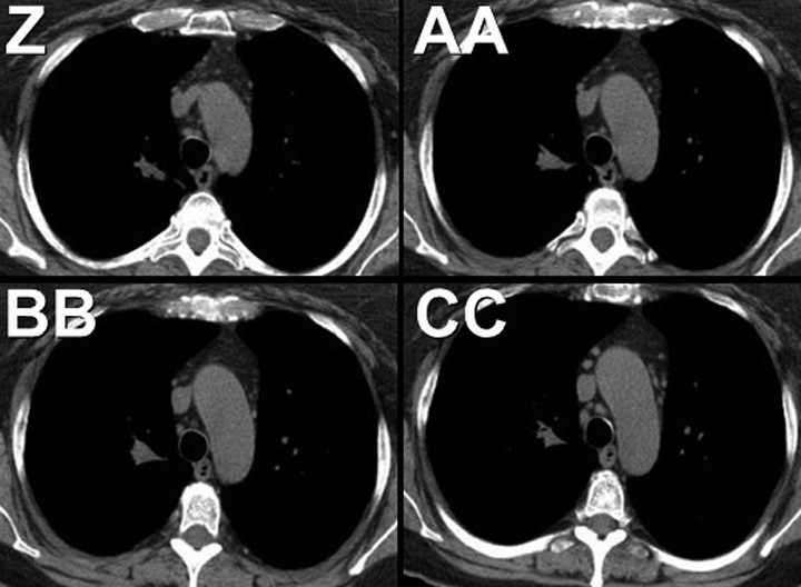
Figure 5. Unenhanced axial thoracic CT displayed in soft tissue windows shows no evidence of calcification within the right apical lesion.
At this point, which of the following represents the most appropriate step in this patient’s management? (Click on the correct answer to be directed to the third of nine pages)