
Correct!
4. The chest radiograph shows a possible focal air space opacity
The frontal chest radiograph shows focal left lung opacity, consistent with air space opacity, with preservation of the left cardiac border. No lung nodules, cavitary or otherwise, are present. The cardiomediastinal contours appear normal. No evidence of interlobular septal thickening is seen. The opacity seen on the chest radiograph is focal, not multifocal, and no pleural effusion is present.
The patient underwent thoracic CT (Figure 2) for further investigation of the chest radiographic findings.
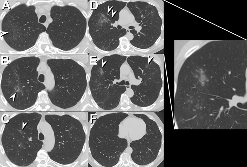
Figure 2. Representative images form the axial thoracic CT performed several weeks after initial presentation, displayed in lung windows, shows patchy, multifocal ground-glass opacity (arrowheads) bilaterally, more pronounced on the right.Relatively little left-sided lung opacity is seen, compared with the chest radiograph performed about 3 weeks earlier. Right: video of thoracic CT scan in lung windows.
Which of the following represent appropriate differential diagnostic considerations for the chest radiographic pattern present? (Click on the correct answer to proceed to the third of nine pages)