
Correct!
1. Left lung atelectasis
His chest x-ray shows rapidly increasing density in the left chest with left lung volume loss. The tip of the endotracheal tube is visible in the trachea. Pleural effusion can cause increased density in the chest but is not associated with volume loss unless there is accompanying atelectasis. His CXR about 12 hours after his intubation is shown in Figure 2.
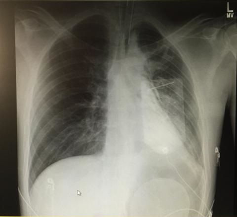
Figure 2. Chest x-ray the next morning after being on volume control ventilator settings overnight. The rapid clearing of the density on the right is consistent with atelectasis.
After transfer to the ICU, his neurologic exam was significant for inability to open his eyes bilaterally, blurred vision (after manual opening of his eyelids), dilated but reactive pupils (8 mm) and symmetric, bilateral, proximal muscle weakness with intact sensation but absent reflexes. He was able to move all 4 extremities spontaneously and could communicate by writing.
Which diagnosis is least likely given the presentation? (Click on the correct answer to proceed to the third of four panels)