
Correct!
5. All of the above
There is a residual pneumothorax from the open lung biopsy, a chest tube present, diffuse ground glass opacities and inter- and intralobular septal thickening (Figure 2).
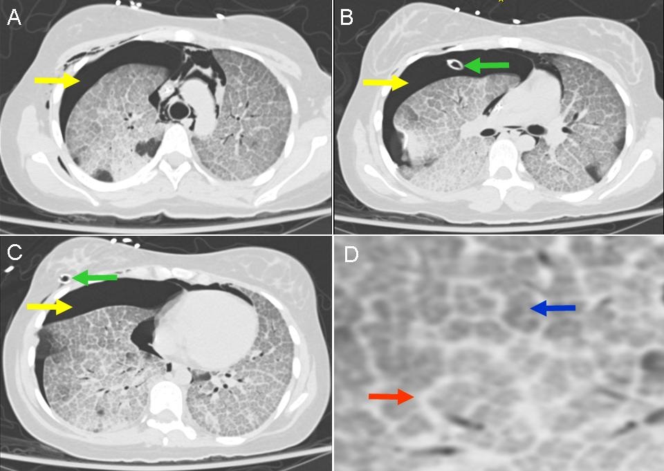
Figure 2. Panels A-C: Thoracic CT scan showing pneumothorax (yellow arrows) and chest tube (green arrows). Panel D: enlargement of the right lung showing interlobular septal thickening (red arrow) and intralobular septal thickening (blue arrow).
The lung biopsy performed at the outside hospital was requested (Figure 3).
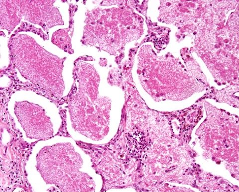
Figure 3. High power view of the open lung biopsy from the outside material showing positive periodic acid-Schiff (PAS) material in the alveoli.
What is the diagnosis? (Click on the correct answer to procced to the third of five panels)