
Correct!
4. 1 and 3
Her ECG shows an irregular rhythm with a rapid ventricular response. There are no consistent P waves proceeding the QRS complex consistent with atrial fibrillation. She was given 10 mg of IV metropolol and her rate decreased to 100-110 and became regular.
Neither diaphragm is identifiable on the portable chest x-ray and there appears to be volume loss with atelectasis. This suggests bilateral pleural effusions.
She is awake, able to follow commands and probably able to protect her airway. CPAP 7 cm H2O, 30% was applied with some improvement.
She was transfused with 2 units of packed red blood cells and had a right-sided thoracentesis which removed 1 liter of straw-colored fluid. After removal of the fluid she had increasing shortness of breath. A lung ultrasound was performed (Figure 3). Lung sliding was seen on both the left and right side.
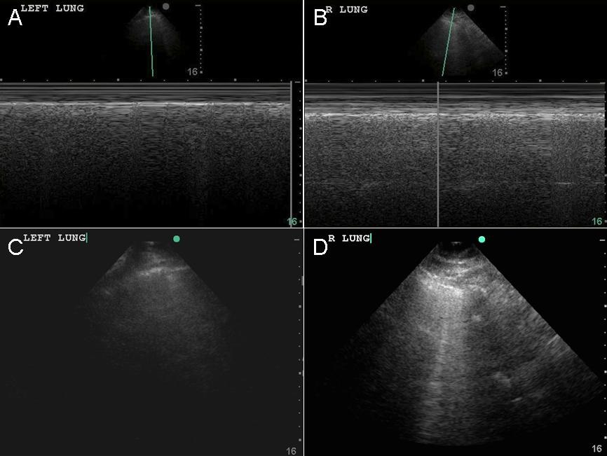
Figure 3. M mode of left lung (panel A) and the right lung (panel B). Ultrasound of left lung (panel C) and right lung (panel D).
Which of the following are true? (Click on the correct answer to proceed to the third of four panels)