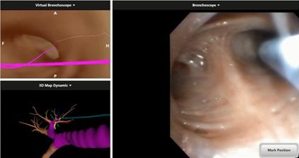
Figure 1. During the first navigation virtual bronchoscope image and 3D map (top left and bottom left) show the tip of the locatable guide in the posterior segment of the right upper lobe matching live video bronchoscope image.
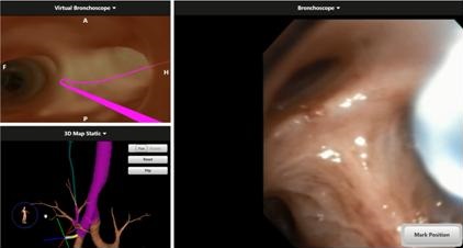
Figure 2. Second navigation: the virtual bronchoscope image and 3D map (top left and bottom left) show the tip of the bronchoscope in the right main bronchus whereas the video bronchoscope shows the tip in the posterior segment of the right upper lobe.
A 59 year-old woman with a 40 pack-year smoking history was referred to our practice with a 2.5 cm spiculated right upper lobe lung nodule for a diagnostic bronchoscopy.
We performed electromagnetic navigation bronchoscopy under general anesthesia in the operating room. After successfully navigating to the lesion and obtaining 3 needle biopsy samples and two cytology brush samples we lost target alignment. After attempting to rotate and reposition the catheter several times it was decided to re-navigate from the trachea. Two images comparing virtual navigation to real anatomy during the first and second navigation attempts are provided bellow (Figures 1 and 2).
Why are the virtual images different? (Click on the correct answer for a discussion)
Cite as: Vazquez-Guillamet R, Horn E, Sarver R, Melendres L. Medical image of the week: virtual anatomical dissociation during electromagnetic navigation bronchoscopy. Southwest J Pulm Crit Care. 2015;11(5):238-9. doi: http://dx.doi.org/10.13175/swjpcc111-15 PDF