Michael B. Gotway, MD
Department of Radiology
Mayo Clinic Arizona
Scottsdale, AZ
Clinical History: A 34-year-old non-smoking woman presented to her physician as an outpatient with complaints of intermittent chest pain and intermittent mild hemoptysis. Her previous medical history was otherwise unremarkable.
Frontal chest radiography (Figure 1) was performed.
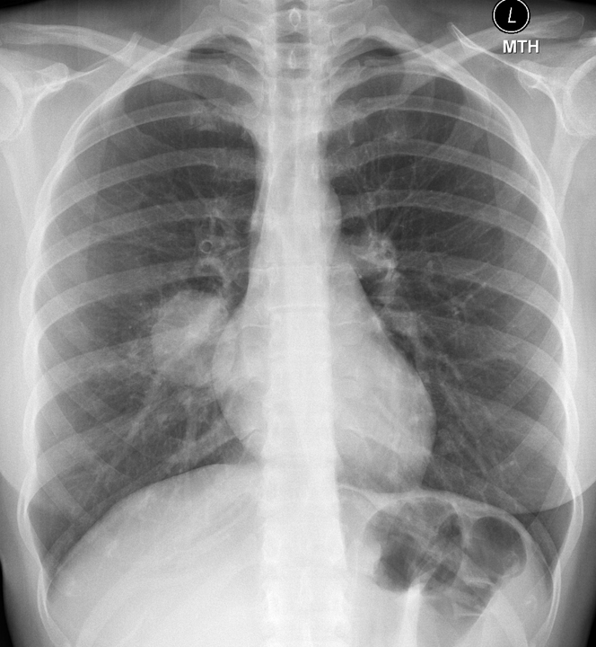
Figure 1. Frontal chest radiography.
Which of the following statements regarding the chest radiograph is most accurate? (Click on the correct answer to proceed to the second of 6 panels)
- The chest radiograph shows a circumscribed pulmonary mass
- The chest radiograph shows asymmetric pulmonary vascularity
- The chest radiograph shows bilateral linear and reticular opacities and diminished lung volumes suggesting fibrotic lung disease
- The chest radiograph shows mild streaky central opacities, possibly reflecting airway thickening
- The chest radiograph shows numerous small nodules
Reference as: Gotway MB. December 2014 imaging case of the month. Southwest J Pulm Crit Care. 2014;9(6):311-9. doi: http://dx.doi.org/10.13175/swjpcc157-14 PDF