Michael B. Gotway, MD
Department of Radiology
Mayo Clinic Arizona
Scottsdale, AZ
Clinical History
A 77-year-old man presented for an executive health physical. His past medical history was significant for coronary artery disease, renal stones, gout, and a left nephrectomy for clear cell renal carcinoma 17 years earlier. Chest radiography (Figure 1) was performed. Prior chest radiographs from the previous year (Figure 2) and 7 years earlier (Figure 3) are shown for comparison.
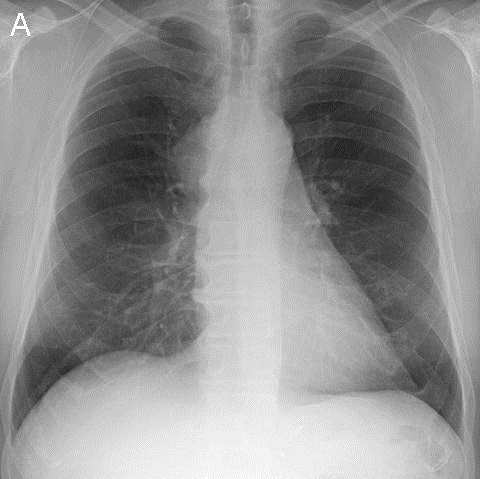
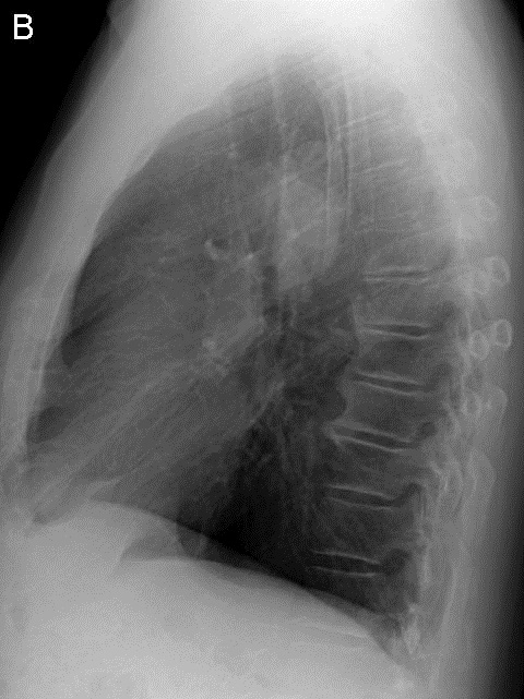
Figure 1. Frontal (A) and lateral (B) chest radiography.
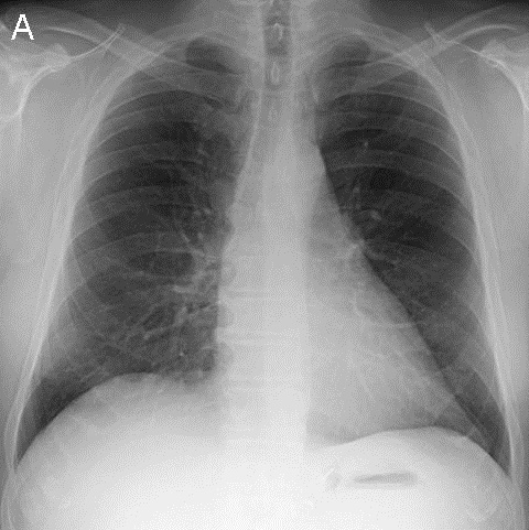

Figure 2. Frontal (A) and lateral (B) chest radiography performed one year prior to presentation.

Figure 3. Frontal chest radiography performed 7 years prior to presentation.
Which of the following statements regarding the chest radiograph is most accurate? (Click on the correct answer to move to next panel)
- The chest radiograph shows a mass
- The chest radiograph shows an unusual cardiac configuration
- The chest radiograph shows basal predominant linear opacities suggesting fibrosis
- The chest radiograph shows multifocal ground-glass opacity and consolidation associated with linear and reticular abnormalities
- The chest radiograph shows multiple nodules
Reference as: Gotway MB. January 2014 imaging case of the month. Southwest J Pulm Crit Care. 2014;8(1):27-40. doi: http://dx.doi.org/10.13175/swjpcc002-14 PDF