
Correct!
1. The chest x-ray shows a mass
The chest radiograph shows a mass along the right mediastinum at the level of the thoracic aortic arch (Figure 4).
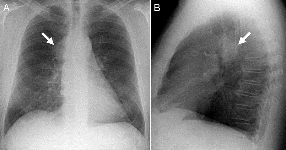
Figure 4. Frontal (A) and lateral (B) chest radiography shows a smoothly contoured, finely lobulated mass along the right paratracheal region (arrow, A), overlying the posterior aortic arch in the lateral projection (arrow, B).
On the lateral radiograph, the mass projects over the posterior aortic arch, suggesting a middle mediastinal location. The lesion is new from the remote prior chest radiograph (Figure 3) and new or at least increasing from one year previously (Figure 2). The lungs appear clear- no nodules, areas of ground-glass opacity, or consolidation are evident, nor are there linear or reticular abnormalities or features to suggest fibrotic lung disease. The heart size and configuration appear normal.
Which of the following is the least appropriate consideration among the differential diagnostic possibilities for the appearance of the patient’s chest radiograph? (Click on the correct answer to move to next panel)