
Correct!
4. 1 or 3
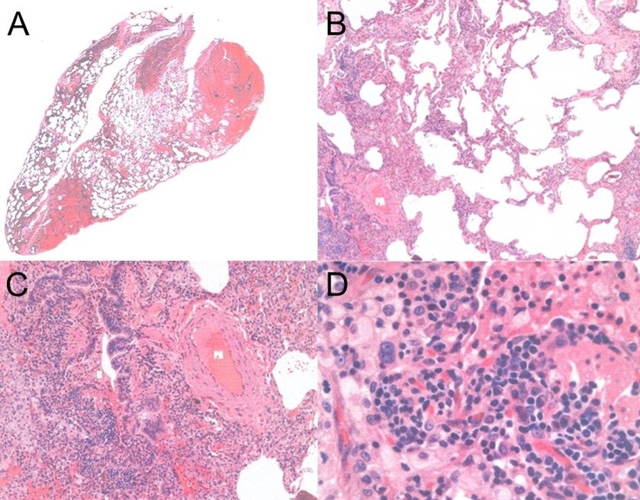
In our experience, a repeat bronchoscopy is rarely diagnostic when the first bronchoscopy does not provide a specific diagnosis. The patient was reluctant to undergo VATS and chose to seek a second opinion. A consulting pulmonologist also recommended VATS which was performed. The biopsy showed patchy interstitial pneumonia and associated chronic broncholitis (Figure 2).

Figure 2. Histology of VATS lung biopsy. A: Low power view demonstrating patchy consolidation with adjacent interstitial infiltrates. B: Intermediate magnification showing an inflammatory process accentuating the centrilobular zone. PA=pulmonary artery. C: Higher magnification showing a prominent chronic inflammatory population admixed with vacuolated macrophages. D: High power magnification of the inflammatory infiltrate showing plasma cells, lymphocytes and aggregates of macrophages. To view Figure 2 in a separate, enlarged window click here.
The pathologist thought the biopsy was most consistent with hypersensitivity pneumonitis (HP) (3).
What should be done next? (Click on the correct answer to be directed to the sixth and final page.)